. 2008 Oct 28;18(20):1537-45.
doi: 10.1016/j.cub.2008.08.033. Epub 2008 Sep 4.

Large ventral lateral neurons modulate arousal and sleep in Drosophila

Affiliations

Large ventral lateral neurons modulate arousal and sleep in Drosophila

Vasu Sheeba et al. Curr Biol. .

Abstract

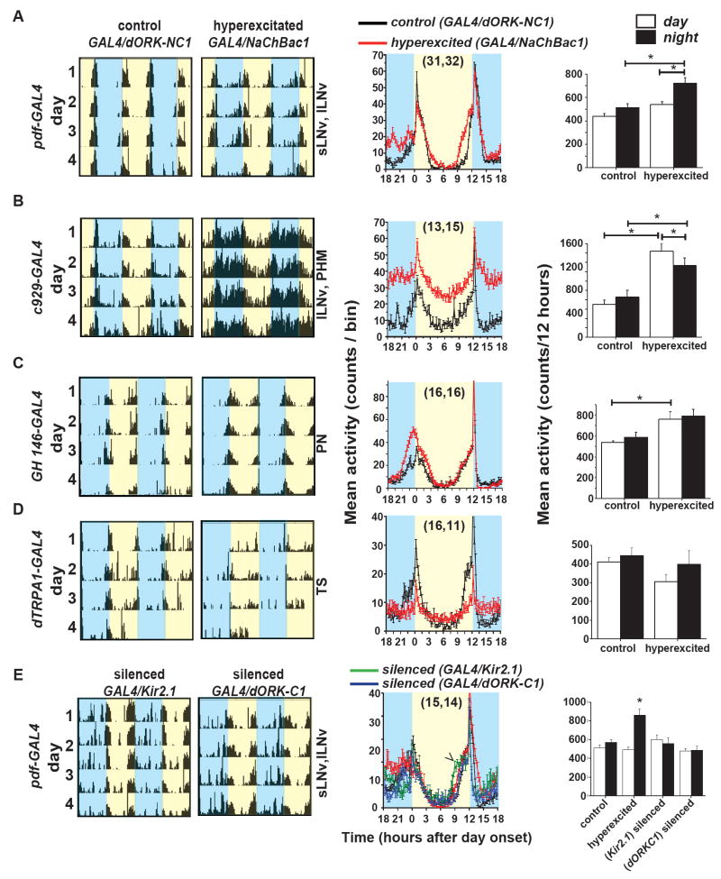
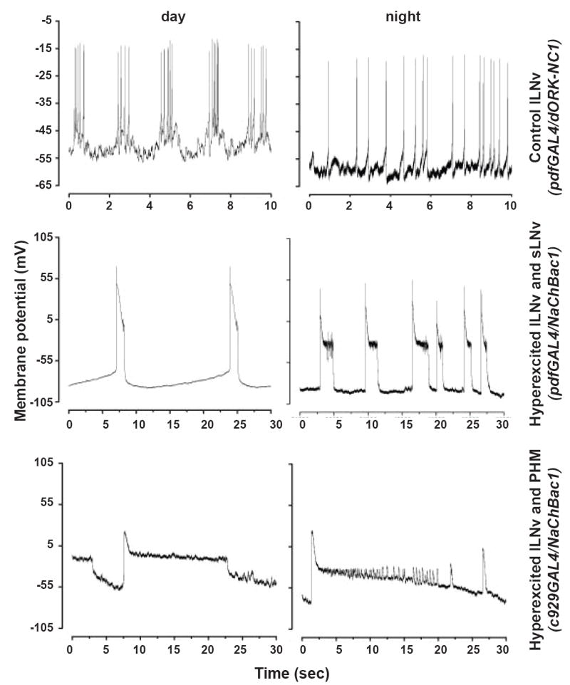
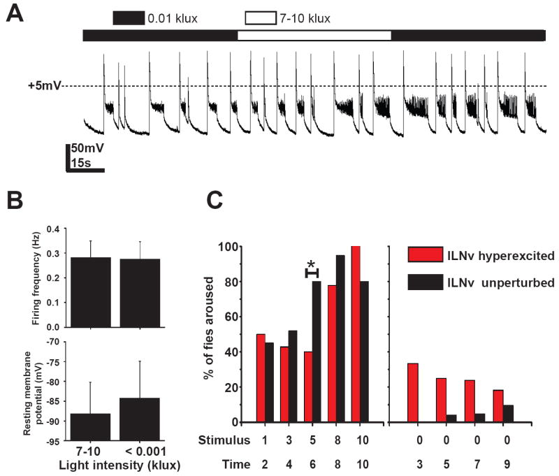
Background: Large ventral lateral clock neurons (lLNvs) exhibit higher daytime-light-driven spontaneous action-potential firing rates in Drosophila, coinciding with wakefulness and locomotor-activity behavior. To determine whether the lLNvs are involved in arousal and sleep/wake behavior, we examined the effects of altered electrical excitation of the LNvs.

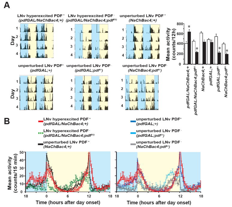
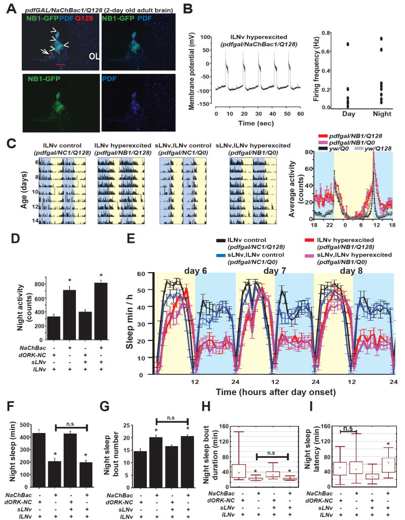
Results: LNv-hyperexcited flies reverse the normal day-night firing pattern, showing higher lLNv firing rates at night and pigment-dispersing-factor-mediated enhancement of nocturnal locomotor-activity behavior and reduced quantity and quality of sleep. lLNv hyperexcitation impairs sensory arousal, as shown by physiological and behavioral assays. lLNv-hyperexcited flies lacking sLNvs exhibit robust hyperexcitation-induced increases in nocturnal behavior, suggesting that the sLNvs are not essential for mediation of arousal.

Conclusions: Light-activated lLNvs modulate behavioral arousal and sleep in Drosophila.


Figures

Figure 1
Figure 1. Behavioural screen of hyperexcitation in different subsets of the circadian pacemaker circuit shows hyperexcitation of lLNv is sufficient for induction of higher nocturnal locomotor activity under 12:12 LD cycles
(A-D) Representative double-plotted actograms of control (dORK-NC1 channel) and LNv hyperexcited (expressing NaChBac1 channel) flies under the control of different GAL4 drivers. Blue shaded areas denote night while yellow shaded areas denote day. The subsets of brain neurons targeted by each of the GAL4 driver lines are indicated next to the actograms. Panels in the middle are the corresponding average profiles of mean activity levels binned across 15 mins. Black lines represent mean activity (± S.E.M) of controls and red lines are hyperexcited flies. Numbers of flies whose activity was used for analysis are in parentheses (GAL4/dORK-NC1, GAL4/Nachbac1). Panels on the right are mean (± S.E.M) day (white) and night (black) activity (counts/12h) for each genotype. Non-parametric analyses using the Kruskal-Wallis ANOVA by ranks followed by multiple comparisons, or one-way ANOVA followed by Tukey’s HSD test were used to determine significant differences, p < 0.05 are indicated by horizontal lines and asterisk. (A) pdf-GAL4 driver targets sLNv and lLNv. (B) c929-GAL4 targets lLNv in addition to nearly 300 peptidergic neurons. (C) GH146-GAL4 does not drive expression in any known circadian neurons and targets projection neurons (PN) in antennal lobe. (D) Temperature sensing (TS neurons) are targeted by dTRPA-GAL4 driver. (E) Flies expressing Kir2.1 (green) and dORK-C1 (blue) ion channels that cause electrical silencing or hypoexcitation of LNv when driven using pdfGAL4 do not show any change in nocturnal activity levels compared to controls but have significantly lower activity than NaChBac1 flies. Kir2.1 expression in the LNv (green line) causes phase advance in the evening peak (arrowhead) and disrupts anticipation in the morning activity peak.
Figure 2
Figure 2. NaChBac expression in LNv causes hyperexcitation and reversal of day/night pattern of net excitation in lLNv
Representative traces of whole cell current clamp recordings from control (uppermost panels, expressing dORK-NC1) or hyperexcited lLNv (middle and lowermost panel, expressing NaChBac1) recorded from brains dissected during the day (left panels, ZT 1-6) or night (right panels, ZT 18-22). At night four out of six control lLNv show tonic firing with mean frequency 1.7 ± 0.5 Hz (mean ± SEM) and resting membrane potential −45 ± 3 mV (the remaining two silent lLNv showed membrane potential within the range of the other four lLNv). Hyperexcited lLNv show large spontaneous depolarisations with mean firing frequency of 0.17 ± 0.04 Hz starting from hyperpolarized resting potentials of −77 ± 14 mV (n = 5) when no other cells in the brain express NaChBac. When NaChBac is expressed using the c929GAL4 driver both day and night phases show higher probability of depolarisation with mean resting membrane potential of −78 ± 5 mV (n = 4 for day and night). (Note the differences in scales of both x and y-axes between uppermost control panels and experimental panels due to extremely large depolarisations in hyperexcited lLNv.
Figure 3
Figure 3. LNv hyperexcitation disrupts nocturnal sleep
(A) Average amount of sleep plotted for days 5, 7, 9 and 11 under 12:12 LD. Error bars are S.E.M. LNv hyperexcited flies (red line) experience significantly lower levels of sleep throughout the night phase compared to the pdfGAL4 driver (grey line) and controls expressing non-conducting dORK-NC1 channel (black line). (B) Mean night time sleep (± SEM) of LNv hyperexcited flies is significantly lower than all other genotypes (One way ANOVA, F4,74 = 14.17, p < 0.001; and post-hoc multiple comparisons with Tukey’s HSD, p < 0.001). (C) Box plot of night time intensity of activity showing LNv hyperexcited flies are not hyperactive relative to the both genetic background controls and dORK-NC1 expressing flies (Kruskal-Wallis Test: H4,80 = 4.7, p = 0.31 (D) Box plot of night time sleep bout duration showing LNv hyperexcited flies experience significantly lower duration sleep episodes relative to the both genetic background controls and dORK-NC1 expressing flies (Kruskal-Wallis Test: H4,77 = 24.3, p <0.001; multiple comparisons, p < 0.01). Median, 25-75 percentile and range are indicated. (E) Sleep latency after night onset is greater in LNv hyperexcited flies compared to all other genotypes (one-way ANOVA F4,75 = 14.5, p < 0.001; Tukey’s HSD, p < 0.05).
Figure 4
Figure 4. Enhanced nocturnal activity behaviour in LNv hyperexcited flies is PDF dependent
(A) Left panel shows representative actograms of LNv hyperexcitation in wild type and pdf01 backgrounds and the control driver line in pdf01 background showing LNv hyperexcitation induced enhanced nocturnal activity only in presence of PDF. Right panel gives mean (± SEM) of activity (counts/12h) during the day (white bar) and night (black bar). * indicates significantly higher night activity count of NaChBac expressing flies in presence of PDF compared to all other genotypes. * also indicates significantly lower activity of pdf01 background flies compared to wild type genetic background. (B, Left panel) Average activity profiles show that hyperexcitation of LNv by NaChBac expression cannot elicit nocturnal behaviour in the absence of PDF (green line). Black line shows wild type activity profile of the NaChBac4 line in the absence of the driver. Red line shows enhanced nocturnal activity when LNv are hyperexcited (pdfGAL4/NaChBac4) in a wild type genetic background. (B, Right panel) Background control activity profiles show profile of the pdfGAL4 driver in a pdf01 genetic background (cyan) with no anticipation of day onset and phase advanced anticipation to night onset, both characteristic features of pdf01 flies. Normal activity profile is seen in the driver line in PDF+ background (dark blue). Both overall activity level and the morning peak is reduced in the absence of PDF (grey and cyan lines) thus PDF in LNv is essential for the overall activity level and the acute response to day onset.
Figure 5
Figure 5. Large LNv drives arousal and modulate sleep independent of functional sLNv
(A) GFP tagged NaChBac1 (Green), anti-PDF (Blue) and anti-HTT (red) staining of 2 day old adult brains of flies co-expressing NaChBac1 and HTT-Q128 (pdfgal/NaChBac/Q128). pdfGAL4 driven NaChBac-GFP and PDF are clearly seen in four lLNv while there is no NaChBac-GFP or PDF in the region where Q128 ablated sLNv are expected to be located. Scale bar = 20 mm. Arrowheads indicate lLNv and arrow points to expected location of sLNv in the accessory medulla. OL = Optic lobe. (B Left panel) Whole cell current clamp recordings during night (ZT 18-22) of lLNv co-expressing NaChBac1 and HTT-Q128 shows typical NaChBac features. (B Right panel) Scatter plot of day and night firing frequencies. (C) Representative actograms showing increased activity during the night in NaChBac expressing flies even when Q128 HTT ablates sLNv. (D) Mean night time locomotor activity is significantly higher when lLNv are hyperexcited both in absence or presence of hyperexcited sLNv (One-way ANOVA F3,121 =33.8, p < 0.001; multiple comparisons p < 0.001 for comparisons between genotypes indicated by * with all others except among themselves). (E) Profile of day and night time sleep under 12:12 LD of flies with hyperexcited lLNv alone (red line, NaChBac1/Q128, n = 30) and both-LNv hyperexcited (pink, NaChBac1/Q0, n = 32) showing decreased nocturnal sleep relative to respective controls where dORK-NC1 is co expressed with HTT-Q128 or Q0 (black, blue; n = 32 for both genotypes). (F) Mean night time sleep is significantly lower when lLNv alone (NaChBac/Q128) or when both sLNv and lLNv (NaChBac/Q0) are hyperexcited compared to controls (dORK-NC1) when either lLNv alone or both LNv are present (One-way ANOVA F3,121 =65.8, p < 0.001; multiple comparisons p < 0.001 for comparisons between genotypes indicated by * with all others except among themselves). (G) The number of night time sleep bouts is significantly higher when lLNv alone or both LNv are hyperexcited (one-way ANOVA F3,120 = 13.2, p < 0.001; Tukey’s HSD, p < 0.02 for comparisons between genotypes indicated by * with all others except among themselves). (H) Duration of the bouts of night time sleep is also significantly shorter when lLNv alone or both LNv are hyperexcited, thus quality of sleep is also significantly affected by selectively hyperexciting lLNv (Kruskal-Wallis Test: H3,124 = 66.3, p < 0.001; multiple comparisons, p < 0.001 for comparisons between genotypes indicated by * with all others except among themselves). (I) The latency of night time sleep of lLNv hyperexcited flies is not significantly greater than control flies with unperturbed lLNv alone while hyperexcitation of both LNv significantly increases sleep latency (Kruskal-Wallis Test: H3,125 = 33.1, p < 0.001; multiple comparisons, p < 0.001).
Figure 6
Figure 6. Membrane hyperexcitation impairs lLNv mediated multisensory input
(A) Representative trace of whole cell current clamp recordings from hyperexcited lLNv in the absence of sLNv, recorded from brains dissected during the night showing no response to a 1000-fold change in light intensity. (B) Bar graphs show no change in firing frequency or resting membrane potential between the two recording conditions (mean ± SEM). (C) Hyperexcitation of lLNv reduces the sensitivity of flies to mechanical stimulation. X- axis top line denotes increasing strength of vertical mechanical stimulation (arbitrary units). The lower line values denote the time of application of stimulus in hours after onset of night. The threshold for arousal in lLNv hyperexcited flies is greater than controls as shown by the left panel where significantly lower fraction of flies can be woken up from sleep at the intermediate stimulus intensity. Right panel shows that LNv hyperexcited flies have an intrinsically higher probability of being awake during the time of the assay compared to controls.

References

    1. Pace-Schott EF, Hobson JA. The neurobiology of sleep: genetics, cellular physiology and subcortical networks. Nature Reviews. 2002;3:591–605. - PubMed
    1. Holmes TC, Sheeba V, Mizrak D, Rubovszky B, Dahdal D. Circuit-breaking and behavioral analysis by molecular genetic manipulation of neural activity in Drosophila. In: North G, Greenspan R, editors. Invertebrate Neurobiology. Cold Spring Harbor: Cold Spring Harbor Laboratory Press; 2007. pp. 19–52.
    1. Sheeba V, Gu H, Sharma VK, O’Dowd DK, Holmes TC. Circadian- and light-dependent regulation of resting membrane potential and spontaneous action potential firing of Drosophila circadian pacemaker neurons. Journal of Neurophysiology. 2008;99:976–988. - PMC - PubMed
    1. Sheeba V, Sharma VK, Gu H, Chou YT, O’Dowd DK, Holmes TC. Pigment dispersing factor-dependent and -independent circadian locomotor behavioral rhythms. J Neurosci. 2008;28:217–227. - PMC - PubMed
    1. Luan H, Lemon WC, Peabody NC, Pohl JB, Zelensky PK, Wang D, Nitabach MN, Holmes TC, White BH. Functional dissection of a neuronal network required for cuticle tanning and wing expansion in Drosophila. J Neurosci. 2006;26:573–584. - PMC - PubMed

Publication types

LinkOut - more resources