Review
. 2008:98:85-120.
doi: 10.1016/S0065-2776(08)00403-3.

New insights on mast cell activation via the high affinity receptor for IgE

Affiliations
Review

New insights on mast cell activation via the high affinity receptor for IgE

Juan Rivera et al. Adv Immunol. 2008.

Abstract

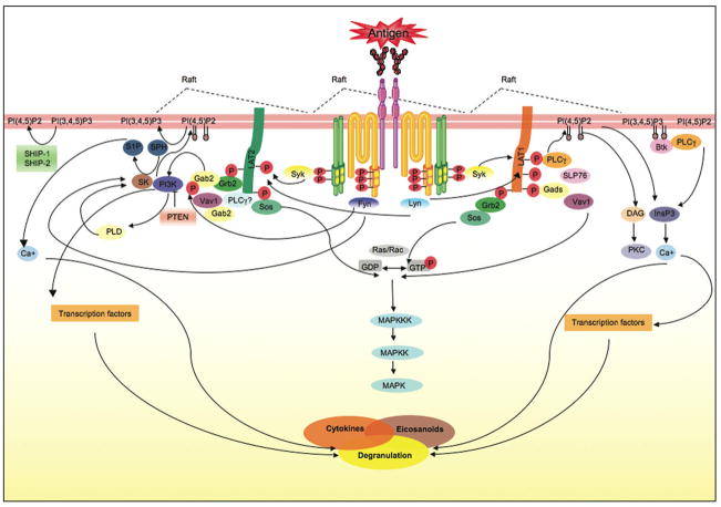
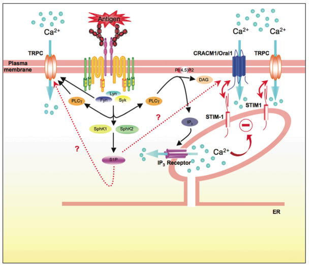
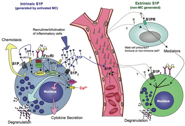
Mast cells are innate immune cells that function as regulatory or effector cells and serve to amplify adaptive immunity. In adaptive immunity these cells function primarily through cell surface Fc receptors that bind immunoglobulin antibodies. The dysregulation of their adaptive role makes them central players in allergy and asthma. Upon encountering an allergen (antigen), which is recognized by immunoglobulin E (IgE) antibodies bound to the high affinity IgE receptor (FcepsilonRI) expressed on their cell surface, mast cells secrete both preformed and newly synthesized mediators of the allergic response. Blocking of these responses is an objective in therapeutic intervention of allergic diseases. Thus, understanding the mechanisms by which antigens elicit mast cell activation (via FcepsilonRI) holds promise toward identifying therapeutic targets. Here we review the most recent advances in understanding antigen-dependent mast cell activation. Specifically, we focus on the requirements for FcepsilonRI activation, the regulation of calcium responses, co-stimulatory signals in FcepsilonRI-mediated mast cell activation and function, and how genetics influences mast cell signaling and responses. These recent discoveries open new avenues of investigation with therapeutic potential.


Figures

Figure 1
Figure 1. FcεRI signaling in mast cells
Antigen-clustering of FcεRI through bound antigen-specific IgE initiates mutiple events required for mast cell activation. Clustering of FcεRI drives the coalesence of cholesterol-enriched membrane microdomains (rafts) that contain signaling molecules like the Src family kinase Lyn. Lyn transphosphorylates the immunoreceptor tyrosine-based activation motifs (ITAMs) of the β and γ subunits of a neighboring FcεRI. This requires that these receptors have an appropriate distance/and or configuration within the antigen-induced receptor clusters. Lyn and Fyn can interact with the phosphorylated β subunits whereas Syk kinase interacts with the γ subunit. Fyn, Lyn, and Syk contribute to the formation of multi-molecular signaling complexes that are coordinated by adaptors, like LAT 1 and 2, Gab2, Grb2, Gads, among others. These signaling complexes provide docking sites for other signaling proteins including PLCγ, SLP76, Vav1, Sos and others. The activity of the molecules in these complexes is coordinated to initiate the production of lipid second messengers that are essential for calcium mobilization and PKC activation leading to degranulation, and the de novo synthesis and secretion of eicosanoids and cytokines. This process is tightly regulated a multiple levels. The signaling complexes provide an environment where a balance of positive signals with negative feedback signals can occur. Lipid enzymes (like PI3K) produce and/or remove lipid messengers (by converting PI(4,5)P2, the substrate of PLCγ, to PI(3,4,5)P3). This production or loss of lipid messengers is important in the targeting, activation and regulation of signaling protein function. Protein kinases can also phosphorylate protein and lipid enzymes modifying their activity. Protein phosphatases (like SHP-1) and lipid phosphatases (like SHIP and PTEN) will dephosphorylate phosphorylated proteins and lipids, respectively, disassembling the signaling complex or inactivating signaling proteins. This is a highly dynamic process that adjusts spatio-temporally to fine-tune mast cell responses.
Figure 2
Figure 2. Calcium Regulation in mast cells
Calcium is an important second messenger whose mobilization is precisely controlled in mast cells. Antigen-aggregation of FcεRI leads to the activation of Lyn, Fyn, and Syk kinases. These kinases contribute to the calcium response by initiating and/or supporting PLCγ activity. PLCγ catalyzes the hydrolysis of membrane-localized phosphatidylinositol 4,5-bisphosphate (PI 4,5-P2) to inositol 3,4,5-trisphosphate (IP3) and diacylglycerol (DAG). IP3 binds IP3 receptors in the ER membrane, resulting in the release of Ca2+ from intracellular Ca2+ stores. DAG regulates the activity of various proteins, such as members of the protein kinase C family. The decrease in the Ca2+ concentration in the ER stores is sensed by STIM-1 (a calcium sensor) causing a change in its conformation that allows its translocation to the plasma membrane where it can interact with CRACM1/Orai1. STIM-1 and CRACM1/Orai1 synergize to elicit the influx of calcium. CRACM1/Orai1 appears to encode the icrac current channel in mast cells originally described by Hoth and Penner. Recent evidence also demonstrates that STIM-1 and Orai1 cooperate with transient receptor potential channels (TRPC) in mast cells to stimulate non-selective entry of Ca2+. These findings demonstrate an increasing complexity in the regulation of Ca2+ entry in mast cells. In other cell types some TRPC’s have also been demonstrated to be targets of Src family kinases, like Fyn, and to be directly regulated by PLCγ. The activation of FcεRI also causes the activation of sphingosine kinases 1 and 2 (SphK1 and SphK2) and the production of sphingosine-1-phosphate (S1P). This sphingolipid regulates Ca2+ influx within the cell, but the intracellular target of S1P is unknown. Moreover the relationship between S1P, STIM-1 and CRACM1/Orai1, if any, remains to be determined.
Figure 3
Figure 3. Role of Sphingosine kinases, S1P and its receptors in mast cell functions
Mast cell function in its physiological environment is affected by “extrinsic S1P” generated by cells other than mast cells (right side, dashed lines with arrows), and “intrinsic S1P” (S1P generated by stimulated mast cells; left side blue arrows). Crosslinking of the FcεRI by IgE/antigen in mast cells results in the rapid activation and translocation of SphK to the plasma membrane and the generation of S1P. This is mediated by the Src kinases Lyn and Fyn. Fyn is required for SphK1 and SphK2 activation, whereas Lyn is required for the early phase of activity and membrane translocation (left panel). Fyn-dependent Gab2/PI3K activation, followed by PLD activation is required for SphK1 stimulation, while a not yet determined Fyn-dependent but Gab2-independent pathway is needed for full SphK2 activation. Our recent studies implicate SphK2, and not SphK1, in the influx of calcium following IgE receptor independently of IP3 generation, and thus affecting mast cell functions. S1P is secreted by activated mast cells to the extracellular media independently of their degranulation via an ATP binding cassette (ABC) transporter (Mitra et al., 2006). Furthermore, S1P is able to rapidly bind and activate its receptors S1P1 and S1P2 on the plasma membrane. S1P1 induces cytoskeletal rearrangements leading to the movement of mast cells towards an antigen gradient, while transactivation of S1P2 enhances the degranulation response. Mast cell secreted S1P can also promote inflammation by activating and recruiting other immune cells involved in allergic and inflammatory responses. Mast cell granules are illustrated as blue circles, and the process of degranulation as granules in contact with the plasma membrane emptying their content (smaller pink and blue dots). Mast cells may be affected by changes in circulating S1P by different mechanisms. It is possible that changes in S1P in the circulation affect the priming of S1P2 in mast cells, which are found in close proximity to blood vessels, enhancing degranulation upon cell activation. Increases or decreases in extrinsic S1P levels might also induce the differentiation of mast cell precursors towards a more or less responsive phenotype, respectively. Constant exposure to higher or lower levels of S1P might also indirectly influence mast cell differentiation or function via mediators derived from other immune or non-immune cells that respond to the fluctuation in S1P levels.

Similar articles

Cited by

References

    1. Ali K, Bilancio A, Thomas M, Pearce W, Gilfillan AM, Tkaczyk C, Kuehn N, Gray A, Giddings J, Peskett E, Fox R, Bruce I, Walker C, Sawyer C, Okkenhaug K, Finan P, Vanhaesebroeck B. Essential role for the p110δ phosphoinositide 3-kinase in the allergic response. Nature. 2004;431:1007–1011. - PubMed
    1. Allende ML, Proia RL. Sphingosine-1-phosphate receptors and the development of the vascular system. Biochim Biophys Acta. 2002;1582:222–227. - PubMed
    1. Allende ML, Sasaki T, Kawai H, Olivera A, Mi Y, van Echten-Deckert G, Hajdu R, Rosenbach M, Keohane CA, Mandala S, Spiegel S, Proia RL. Mice deficient in sphingosine kinase 1 are rendered lymphopenic by FTY720. J Biol Chem. 2004;279:52487–52492. - PubMed
    1. Ammit AJ, Hastie AT, Edsall LC, Hoffman RK, Amrani Y, Krymskaya VP, Kane SA, Peters SP, Penn RB, Spiegel S, Panettieri RA., Jr Sphingosine 1-phosphate modulates human airway smooth muscle cell functions that promote inflammation and airway remodeling in asthma. Faseb J. 2001;15:1212–1214. - PubMed
    1. Baba Y, Nishida K, Fujii Y, Hirano T, Hikida M, Kurosaki T. Essential function for the calcium sensor STIM1 in mast cell activation and anaphylactic responses. Nat Immunol. 2008;9:81–88. - PubMed

LinkOut - more resources