. 2008 Sep 9;14(3):263-73.
doi: 10.1016/j.ccr.2008.08.001.

Rapid chemotherapy-induced acute endothelial progenitor cell mobilization: implications for antiangiogenic drugs as chemosensitizing agents

Affiliations

Rapid chemotherapy-induced acute endothelial progenitor cell mobilization: implications for antiangiogenic drugs as chemosensitizing agents

Yuval Shaked et al. Cancer Cell. .

Abstract

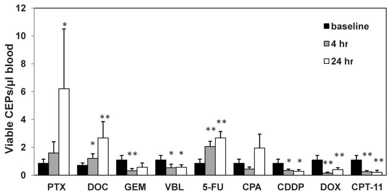
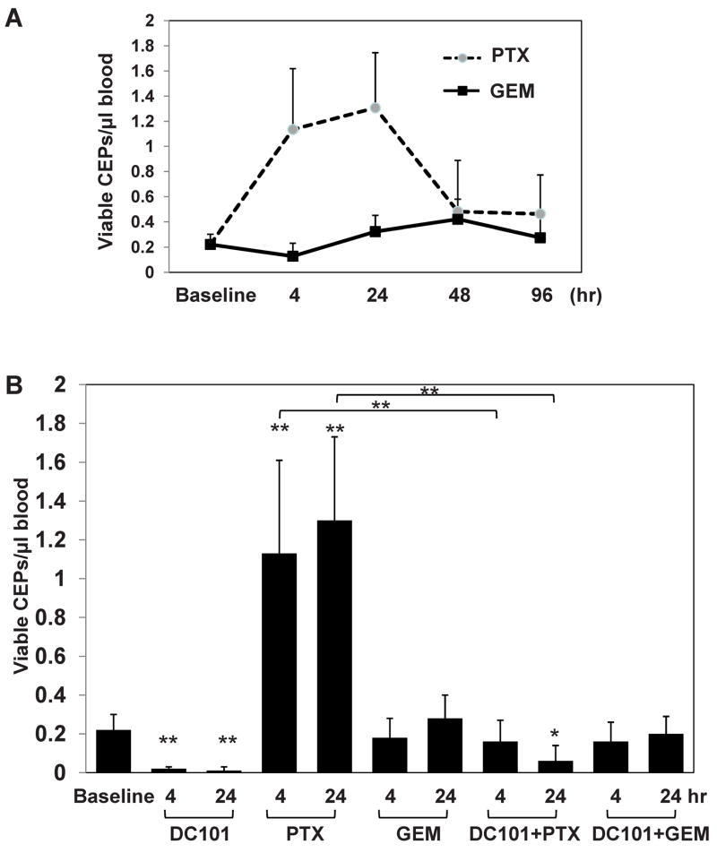
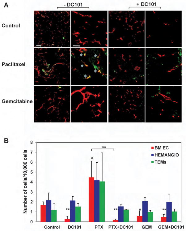
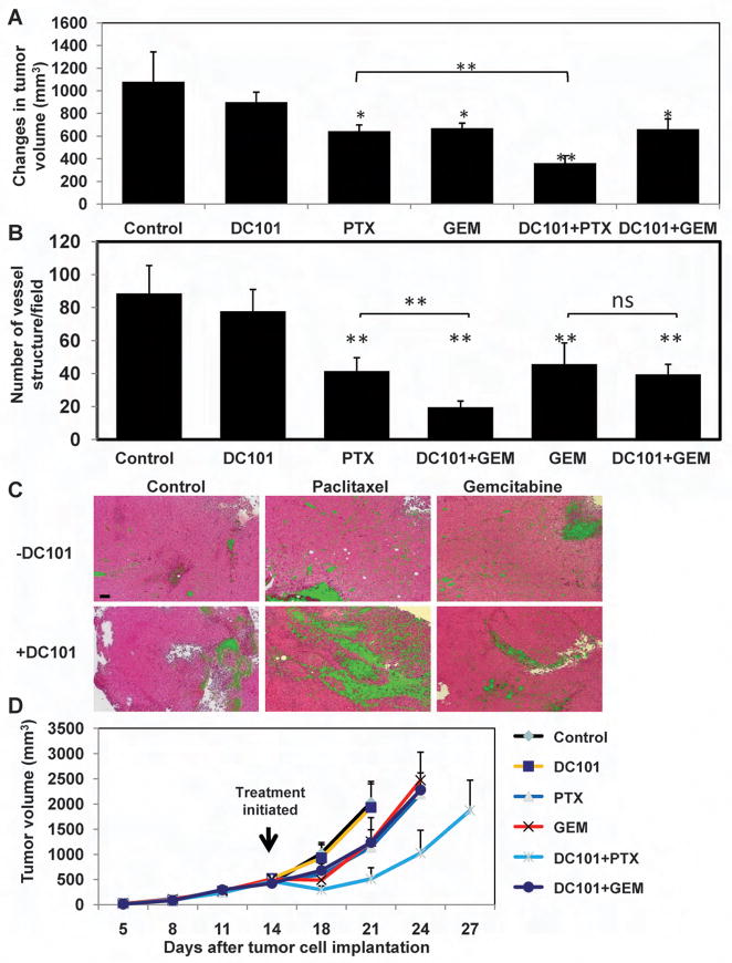
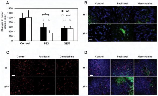
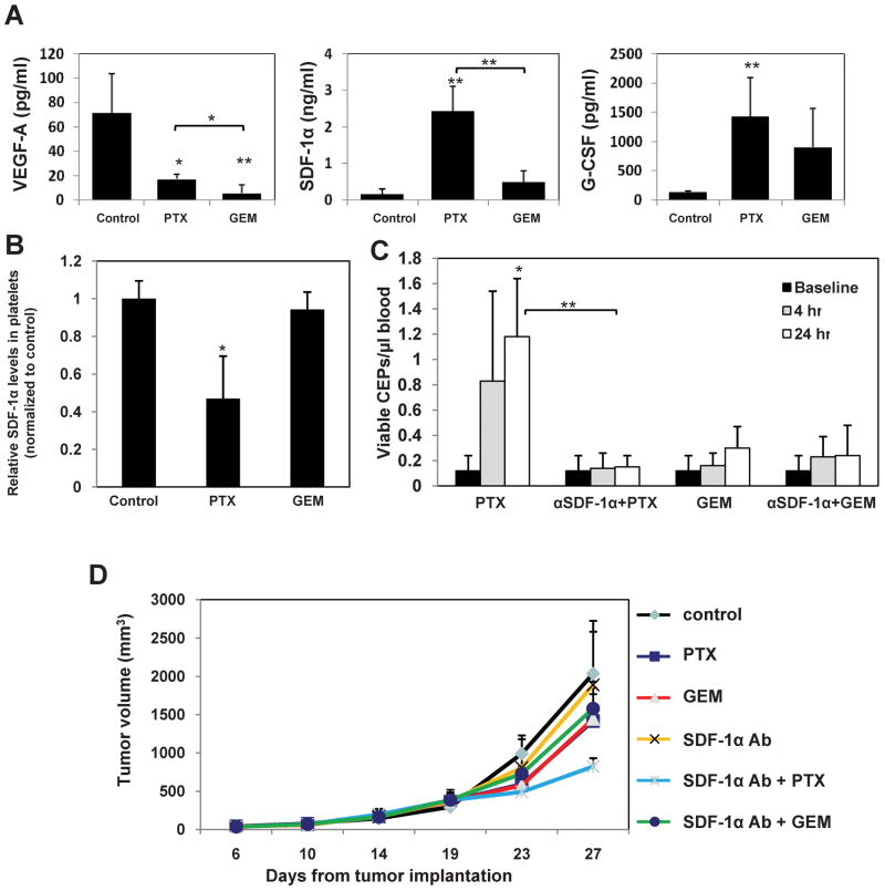
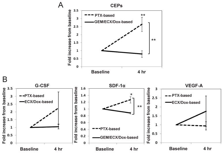
Several hypotheses have been proposed to explain how antiangiogenic drugs enhance the treatment efficacy of cytotoxic chemotherapy, including impairing the ability of chemotherapy-responsive tumors to regrow after therapy. With respect to the latter, we show that certain chemotherapy drugs, e.g., paclitaxel, can rapidly induce proangiogenic bone marrow-derived circulating endothelial progenitor (CEP) mobilization and subsequent tumor homing, whereas others, e.g., gemcitabine, do not. Acute CEP mobilization was mediated, at least in part, by systemic induction of SDF-1alpha and could be prevented by various procedures such as treatment with anti-VEGFR2 blocking antibodies or paclitaxel treatment in CEP-deficient Id mutant mice, both of which resulted in enhanced antitumor effects mediated by paclitaxel, but not by gemcitabine.


Figures

Figure 1
Figure 1. Levels of viable CEPs in non-tumor bearing BALB/c mice treated with a variety of chemotherapy drugs near or at the MTD
8–12 week old BALB/c mice (n=4–5 mice/group) were treated with 30mg/kg paclitaxel (PTX), 120mg/kg gemcitabine, (GEM), 40mg/kg docetaxel (DOC), 11mg/kg vinblastine (VBL), 100mg/kg 5 fluorouracil (5-FU), 250mg/kg cyclophosphamide (CPA), 6mg/kg cisplatinum (CDDP), 12mg/kg doxorubicin (DOX) or 100mg/kg irinotecan (CPT-11) as also indicated in Table S1. Four and 24 hours later mice were bled via retro-orbital sinus for the evaluation of viable CEPs by four color flow cytometry. *, 0.05>p>0.01; **, p<0.01.
Figure 2
Figure 2. Evaluation of CEPs in mice treated with either paclitaxel or gemcitabine in combination with DC101
(A) 8–10 week old non-tumor bearing C57Bl/6 mice (n=4 mice/group) were treated with 50mg/kg paclitaxel (PTX), or 500mg/kg gemcitabine (GEM). Blood was drawn from the retro-orbital sinus at time points indicated in the figure, and processed for the evaluation of viable CEPs using flow cytometry. (B) In a separate experiment, mice were treated with paclitaxel (PTX) or gemcitabine (GEM) as described in (A), with or without DC101 given 24 hours prior to chemotherapy treatment. Blood was drawn via retro-orbital sinus and processed for the evaluation of viable CEPs using flow cytometry. *, 0.05>p>0.01; **, p<0.01.
Figure 3
Figure 3. Homing and colonization of GFP+ bone marrow cells in LLC tumors after treatment with paclitaxel or gemcitabine in combination with DC101
C57Bl/6 mice (n=5 mice/group) that were previously lethally irradiated and subsequently transplanted with 107 GFP+ bone marrow cells obtained from UBI/GFP/C57Bl/6 mice, were used as recipients for a subcutaneous injection of LLC cells which were allowed to grow until they reached 500mm3, at which point treatment with paclitaxel (PTX), gemcitabine (GEM) with or without upfront treatment with DC101 was initiated. Three days later, tumors were removed and sections were prepared for the assessment of (A) GFP+ cells (in green) colonization of the tumors, CD31 staining (in red) as an endothelial cell marker, and blue arrows for colocalization of CD31 and GFP+ cells in the paclitaxel treated group (scale bars left 20μm, right 50μm), or (B) the number of bone marrow derived GFP+ endothelial cells (BM EC), hemangiocytes (Hemangio), and TEMs colonizing the tumor using tumors prepared as a single cell suspension and evaluated by flow cytometry.
Figure 4
Figure 4. Assessment of LLC tumor volume, microvessel density, necrosis, and long-term tumor growth in mice treated with paclitaxel, or gemctibine, in combination with DC101
500mm3 LLC bearing C57Bl/6 mice (n=4–5 mice/group) were treated with paclitaxel (PTX), gemcitabine (GEM) in combination with DC101 administered 24 hours prior to the chemotherapy drug. (A) tumor volumes were assessed before and three days after treatment. The changes in tumor volume are shown. Three days after treatment tumors were removed and evaluated for (B) microvessel density after CD31 staining for vessel structure. Data presented as the number of vessel structures per field (n>10 fields/tumor), or (C) necrosis (in green) on H&E staining (scale bar, 100μm)(see Supplemental Figure S2B for summary of quantitative data). (D) In a separate experiment, LLC tumors implanted in C57Bl/6 mice were allowed to reach 500mm3, at which point treatment with paclitaxel, gemcitabine (administered at the MTDs) and DC101 was initiated. Tumors were measured regularly using a caliper, and tumor growth was plotted as per number of days from tumor cell implantation. *, 0.05>p>0.01; **, p<0.01.
Figure 5
Figure 5. Assessment of LLC tumor volume, hypoxia, perfusion, microvessel density, and tumor cell proliferation and apoptosis of tumors grown in Id mutant mice or their wt controls after treatment with paclitaxel, or gemctibine
500mm3 LLC bearing Id mutant mice were treated with paclitaxel (PTX) or gemcitabine (GEM). (A) tumor volumes were assessed before and three days after treatment. The changes in tumor volume are shown. Three days after treatment tumors were removed and evaluated for (B) vessel perfusion (in blue) and hypoxia (in green) (scale bar, 50μm), (C) microvessel density (CD31 staining in red)(scale bar, 50μm), and (D) proliferation (in red) and apoptosis (in green)(scale bar, 50μm). ns, not significant; *, 0.05>p>0.01; **, p<0.01. See Supplemental Figure S4 for summary of quantitative data, respectively.
Figure 6
Figure 6. Circulating levels of VEGF-A, G-CSF and SDF-1α four hours after treatment with paclitaxel or gemcitabine and the impact of SDF-1α neutralizing antibody treatment on viable CEPs and tumor growth
Non tumor bearing C57Bl/6 mice (n=4 mice/group) were treated with paclitaxel (PTX) or gemcitabine (GEM). Four hours later, mice were bled by cardiac puncture and plasma was collected. (A) Levels of murine VEGF-A, G-CSF and SDF-1α were analyzed by ELISA. (B) Analysis of SDF-1α content stored in isolated circulating platelets from C57Bl/6 mice, 4 hours after they were treated with paclitaxel or gemcitabine at MTDs. (C) Non-tumor bearing C57Bl/6 (n=4–5 mice/group) mice were treated with SDF-1α neutralizing antibodies. Twenty-four hours later, mice were treated with paclitaxel (PTX) or gemcitabine (GEM). After 4 hours, mice were bled from the retro-orbital sinus for the evaluation of viable CEPs by flow cytometry. (D) In C57Bl/6 mice, LLC tumors were allowed to growth until they reached 500mm3, at which point the mice were treated with polyclonal SDF-1α neutralizing antibodies in combination with either paclitaxel or gemcitabine. Control mice received non-specific antisera treatment. Tumors were measured regularly using a caliper, and tumor growth was plotted as per number of days from tumor cell implantation. *, 0.05>p>0.01; **, p<0.01.
Figure 7
Figure 7. Levels of CEPs as well as G-CSF, SDF-1α and VEGF plasma concentrations in cancer patients 4 hours after they were treated with various chemotherapy drugs administered at the MTDs
Cancer patients (n=30) were treated with paclitaxel (n=8), paclitaxel plus carboplatin (n=4)(both of which designated as PTX-based therapy), gemcitabine (n=8)(GEM), epirubicin, cisplatin plus capecitebin (n=5)(ECX), or doxorubicin +/− cyclophosphamide (n=5)(Dox-based therapy). Four hours later, patients were bled intravenously for the evaluation of (A) CEPs (n=12 for PTX-based, and n=18 for GEM/ECX/Dox-based therapies) as well as (B) G-CSF (n=3 for PTX-based, and n=10 for ECX/Dox-based therapies), SDF-1α (n=12 for PTX-based, and n=15 for GEM/ECX/Dox-based therapies) and VEGF (n=3 for PTX-based, and n=10 for ECX/Dox-based therapies) plasma concentrations. Results were normalized to the baseline level of each patient to reduce variability that may occur due to tumor type, stage, and values obtained from two different centers. *, 0.05>p>0.01; **, p<0.01.

Comment in

References

    1. Addison CL, Arenberg DA, Morris SB, Xue YY, Burdick MD, Mulligan MS, Iannettoni MD, Strieter RM. The CXC chemokine, monokine induced by interferon-gamma, inhibits non-small cell lung carcinoma tumor growth and metastasis. Hum Gene Ther. 2000;11:247–261. - PubMed
    1. Asahara T, Takahashi T, Masuda H, Kalka C, Chen D, Iwaguro H, Inai Y, Silver M, Isner JM. VEGF contributes to postnatal neovascularization by mobilizing bone marrow-derived endothelial progenitor cells. EMBO J. 1999;18:3964–3972. - PMC - PubMed
    1. Athanassakis I, Papadimitriou L, Vassiliadis S. Murine ectoplacental cone-derived trophoblast cells express chemokine receptors. J Reprod Immunol. 2001;50:105–119. - PubMed
    1. Avecilla ST, Hattori K, Heissig B, Tejada R, Liao F, Shido K, Jin DK, Dias S, Zhang F, Hartman TE, Hackett NR, Crystal RG, Witte L, Hicklin DJ, Bohlen P, Eaton D, Lyden D, de Sauvage F, Rafii S. Chemokine-mediated interaction of hematopoietic progenitors with the bone marrow vascular niche is required for thrombopoiesis. Nat Med. 2004;10:64–71. - PubMed
    1. Baeten CI, Wagstaff J, Verhoeven IC, Hillen HF, Griffioen AW. Flow cytometric quantification of tumour endothelial cells; an objective alternative for microvessel density assessment. Br J Cancer. 2002;87:344–347. - PMC - PubMed

Publication types

MeSH terms