

doi: 10.1074/jbc.C800160200.
Epub 2008 Sep 4.
Full-length Escherichia coli SecA dimerizes in a closed conformation in solution as determined by cryo-electron microscopy
Affiliations
- PMID: 18772144
- PMCID: PMC2570880
- DOI: 10.1074/jbc.C800160200
Item in Clipboard
Full-length Escherichia coli SecA dimerizes in a closed conformation in solution as determined by cryo-electron microscopy
J Biol Chem.
.
Abstract
SecA is an obligatory component of the Escherichia coli general secretion pathway. However, the oligomeric structure of SecA and SecA conformational changes during translocation processes are still unclear. Here we obtained the three-dimensional structure of E. coli wild-type full-length SecA in solution by single particle cryo-electron microscopy and determined its oligomeric organization. In this structure, SecA occurs as a dimer in which the two protomers are arranged in an antiparallel mode, with a novel electrostatic interface, and both protomers are in closed conformation. The system developed here may provide a promising technique for studying dynamic structural changes in SecA.
Figures
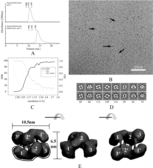
The E. coli SecA structure in solution. A,
size-exclusion chromatography of SecA at 4 °C. A 125-μg sample of SecA
was injected to a Superdex 200 column and eluted in a buffer of 20
mm HEPES-KOH, pH 7.5, or 20 mm HEPES-KOH, 300
mm KCl, pH 7.5. Marker proteins are: Ferritin (440 kDa), bovine
catalase (232 kDa), and aldolase (158 kDa). B, cryo-EM image of 100
μg/ml SecA in buffer (20 mm Tris-Ac, pH 7.5, 1 mm
DTT). Some individual particles are indicated by black arrows. The
bar is 100 nm. C, resolution curves of the three-dimensional
reconstruction. The resolutions calculated from two methods are shown: Fourier
shell correlation (FSC) function (dashed line) and
differential phase residual (DPR) function (solid line). The
dotted line is the critical Fourier shell correlation function
3δ. D, distinct views of SecA cryo-EM samples. The top
panel is the projection map of the reconstructed model; the bottom
panel is the average map of all particles in this class; the number of
particles in this class is indicated in the bottom panel. The
box is 18.4 nm. E, surface representation of the
three-dimensional reconstruction. The surface was rendered and displayed using
the VMD software. Three views are shown: top view, side view, and bottom view.
Each view was obtained after 90° rotation around the horizontal axis, as
shown between the views. The three domains in the two subunits are designated
as 1, 2, and 3, and 1′, 2′,
and 3′.
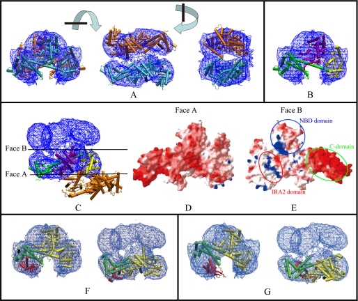
Docking of the cryo-EM three-dimensional map with the ecSecA crystal
structure and comparison of the structures from cryo-EM and X-rays.
A, chain A in the ecSecA (PDB number: 2FSF) was used for docking. The
blue mesh represents the 1.5 σ contour of the electron density
map. There are two molecules in the cryo-EM map, colored cyan and
orange. Three views are shown. Each view is obtained after 90°
rotation around the perpendicular or horizontal axis, as shown between the
views. B, the docked crystal structure is rendered in different
colors for different domains. For clarity, only one SecA protomer is shown.
Similar to the assignment by Papanikolau et al.
(19), ecSecA is divided into
four parts: the NBD domain (purple); the Var, IRA2, and joint domains
(yellow); the PBD domain (red); the C-terminal 30-kDa domain
comprised of the IRA1, WD, SD, and CTD subdomains (green). As noted
above, only a small fraction of PBD structure is seen in the refined x-ray
structure. C, manual docking of one protomer (in different domain
colors) in the x-ray dimeric structure (PDB number: 2FSF) into the electron
density map. The colors in different domains were assigned as in B.
The other protomer (orange) is obviously displaced in the EM map. The
two side surfaces of the SecA protomer are designated Face A and Face B.
D, view of Face A of the SecA protomer after electrostatic surface
calculation. Electrostatic potential is represented as a tricolor gradient
from blue (+1.8 kT (k, the constant of boltzmann;
T, Thermodynamic temperature)) through white (neutral) to
red (–5.8 kT), as represented using the
Swiss-PDBviewer and calculated by DELPHY. E, view of Face B of the
SecA protomer after electrostatic surface calculation. The patches of the
C-domain, the IRA2 domain, and the NBD domain are indicated. The two protomers
interact with each other through this interface. F and G,
docking of the cryo-EM map with bsSecA x-ray structure in the closed (PDB
number: 1M6N) (F) or open (PDB number: 1TF5) (G) state. For
clarity, only one SecA protomer is shown. The PBD domain is red, and
the C-domain is green. Both the side and the top view are shown.
References
-
- Cabelli, R. J., Chen, L., Tai, P. C., and Oliver, D. B. (1988) Cell 55 683–692 - PubMed
-
- Oliver, D. B., and Beckwith, J. (1981) Cell 25 765–772 - PubMed
-
- Manting, E. H., and Driessen, A. J. (2000) Mol. Microbiol. 37 226–238 - PubMed
-
- Wickner, W., and Leonard, M. R. (1996) J. Biol. Chem. 271 29514–29516 - PubMed
Publication types
MeSH terms
Substances
Grants and funding
LinkOut - more resources
Full Text Sources
Molecular Biology Databases
