

Identification of proteins binding to E-Box/Ku86 sites and function of the tumor suppressor SAFB1 in transcriptional regulation of the human xanthine oxidoreductase gene
- PMID: 18772145
- PMCID: PMC2573066
- DOI: 10.1074/jbc.M802076200
Identification of proteins binding to E-Box/Ku86 sites and function of the tumor suppressor SAFB1 in transcriptional regulation of the human xanthine oxidoreductase gene
Abstract
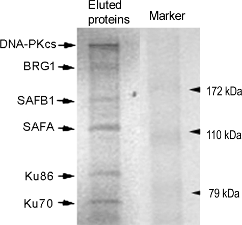
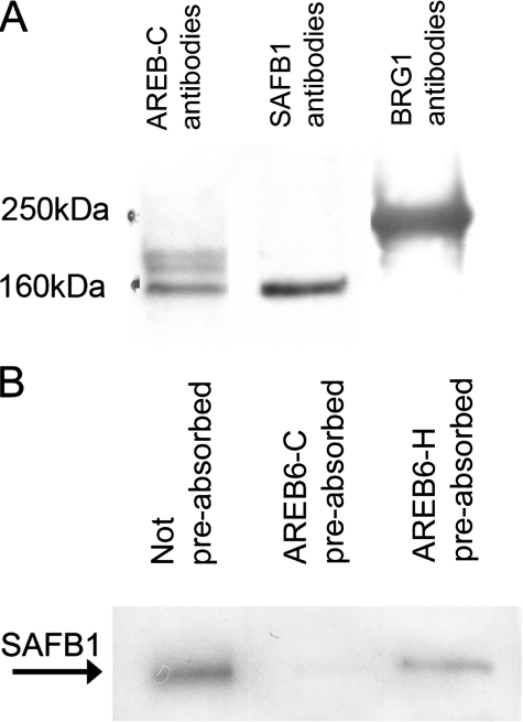
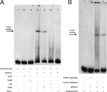
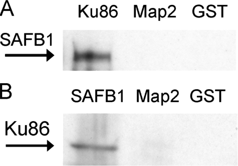
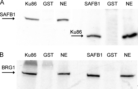
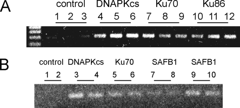
The xanthine oxidoreductase gene (XOR) encodes an important source of reactive oxygen species and uric acid, and its expression is associated with various human diseases including several forms of cancer. We previously reported that basal human XOR (hXOR) expression is restricted or repressed by E-box and TATA-like elements and a cluster of transcriptional proteins, including AREB6-like proteins and DNA-dependent protein kinase (DNA-PK). We now demonstrate that the cluster contains the tumor suppressors SAFB1, BRG1, and SAF-A. We further demonstrate that SAFB1 silencing increases hXOR expression and that SAFB1 directly binds to the E-box. Multiple studies in vitro and in vivo including pulldown, immunoprecipitation and chromatin immunoprecipitation analyses indicate that SAFB1, Ku86, and BRG1 associate with each other. The results suggest that the SAFB1 complex binds to the hXOR promoter in a chromatin environment and plays a critical role in restricting hXOR expression via its direct interaction with the E-box, DNA-PK, and tumor suppressors. Moreover, we demonstrate that the cytokine, oncostatin M (OSM), induces the phosphorylation of SAFB1 and that the OSM-induced hXOR mRNA expression is significantly inhibited by silencing the DNA-PK catalytic subunit or SAFB1 expression. The present studies for the first time demonstrate that hXOR is a tumor suppressor-targeted gene and that the phosphorylation of SAFB1 is regulated by OSM, providing a molecular basis for understanding the role of SAFB1-regulated hXOR transcription in cytokine stimulation and tumorigenesis.
Figures


Similar articles
-
Characterization of proteins binding to E-box/Ku86 sites and function of Ku86 in transcriptional regulation of the human xanthine oxidoreductase gene.J Biol Chem. 2004 Apr 16;279(16):16057-63. doi: 10.1074/jbc.M305856200. Epub 2004 Feb 4. J Biol Chem. 2004. PMID: 14761964
-
Scaffold attachment factor SAFB1 suppresses estrogen receptor alpha-mediated transcription in part via interaction with nuclear receptor corepressor.Mol Endocrinol. 2006 Feb;20(2):311-20. doi: 10.1210/me.2005-0100. Epub 2005 Sep 29. Mol Endocrinol. 2006. PMID: 16195251
-
SAFB1 mediates repression of immune regulators and apoptotic genes in breast cancer cells.J Biol Chem. 2010 Feb 5;285(6):3608-3616. doi: 10.1074/jbc.M109.066431. Epub 2009 Nov 9. J Biol Chem. 2010. PMID: 19901029 Free PMC article.
-
Scaffold attachment factors SAFB1 and SAFB2: Innocent bystanders or critical players in breast tumorigenesis?J Cell Biochem. 2003 Nov 1;90(4):653-61. doi: 10.1002/jcb.10685. J Cell Biochem. 2003. PMID: 14587024 Review.
-
SAFB1- and SAFB2-mediated transcriptional repression: relevance to cancer.Biochem Soc Trans. 2012 Aug;40(4):826-30. doi: 10.1042/BST20120030. Biochem Soc Trans. 2012. PMID: 22817742 Review.
Cited by
-
Xanthine oxidoreductase in cancer: more than a differentiation marker.Cancer Med. 2016 Mar;5(3):546-57. doi: 10.1002/cam4.601. Epub 2015 Dec 21. Cancer Med. 2016. PMID: 26687331 Free PMC article. Review.
-
Acquired xanthine dehydrogenase expression shortens survival in patients with resected adenocarcinoma of lung.Tumour Biol. 2012 Oct;33(5):1727-32. doi: 10.1007/s13277-012-0431-2. Epub 2012 Jun 8. Tumour Biol. 2012. PMID: 22678977
-
DNA-PKcs, a player winding and dancing with RNA metabolism and diseases.Cell Mol Biol Lett. 2025 Mar 4;30(1):25. doi: 10.1186/s11658-025-00703-z. Cell Mol Biol Lett. 2025. PMID: 40038612 Free PMC article. Review.
-
Proteomics-Based Identification of the Molecular Signatures of Liver Tissues from Aged Rats following Eight Weeks of Medium-Intensity Exercise.Oxid Med Cell Longev. 2016;2016:3269405. doi: 10.1155/2016/3269405. Epub 2016 Dec 27. Oxid Med Cell Longev. 2016. PMID: 28116034 Free PMC article.
-
SKA1 promotes tumor metastasis via SAFB-mediated transcription repression of DUSP6 in clear cell renal cell carcinoma.Aging (Albany NY). 2022 Dec 2;14(23):9679-9698. doi: 10.18632/aging.204418. Epub 2022 Dec 2. Aging (Albany NY). 2022. PMID: 36462498 Free PMC article.
References
-
- Massey, V., Brumby, P. E., and Komai, H. (1969) J. Biol. Chem. 244 1682–1691 - PubMed
-
- Waud, W. R., and Rajagopalan, K. V. (1976) Arch. Biochem. Biophys. 172 365–379 - PubMed
-
- Amaya, Y., Yamazaki, K., Sato, M., Noda, K., Nishino, T., and Nishino, T. (1990) J. Biol. Chem. 265 14170–14175 - PubMed
-
- Harrison, R. (2002) Free Radic. Biol. Med. 33 774–779 - PubMed
-
- Knaus, U. G., Heyworth, P. G., Evans, T., Curnutte, J. T., and Bokoch, G. M. (1991) Science 254 1512–1515 - PubMed
Publication types
MeSH terms
Substances
Grants and funding
LinkOut - more resources
Full Text Sources
Research Materials
Miscellaneous
