. 2008 Nov 1;586(21):5077-89.
doi: 10.1113/jphysiol.2008.155127. Epub 2008 Sep 4.

The accessibility and interconnectivity of the tubular system network in toad skeletal muscle

Affiliations

The accessibility and interconnectivity of the tubular system network in toad skeletal muscle

Joshua N Edwards et al. J Physiol. .

Abstract

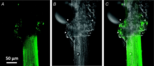
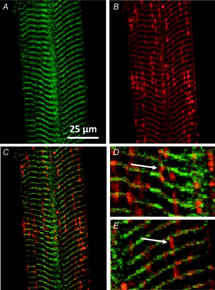
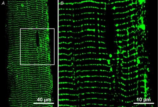
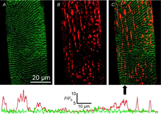
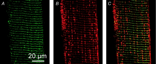
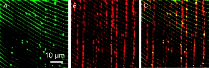
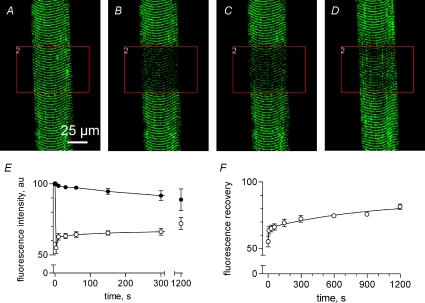
The tubular (t) system is essential for normal function of skeletal muscle fibre, acting as a conduit for molecules and ions within the cell. However, t system accessibility and interconnectivity have been mainly assessed in fixed cells where the t system no longer fully represents that of the living cell. Here, fluorescent dyes of different diameter were allowed to equilibrate within the t system of intact fibres from toad, mechanically skinned to trap the dyes, and then imaged using confocal microscopy to investigate t system accessibility and interconnectivity. Dual imaging of rhod-2 and a 500 kDa fluorescein dextran identified regions throughout the t system that differed in the accessibility to molecules of different molecular weight. Restrictions within the t system lumen occurred at the junctions of the longitudinal and transverse tubules and also where a transverse tubule split into two tubules to maintain their alignment with Z-lines of adjacent mis-registered sarcomeres. Thus, three types of tubule, transverse, longitudinal and Z, can be identified by their lumenal diameter in this network. The latter we define for the first time as a tubule with a narrow lumen that is responsible for the change in register. Stretch-induced t system vacuolation showed exclusive access of rhod-2 to these structures indicating their origin was the longitudinal tubules. Exposing the sealed t system to highly hypertonic solution reversed vacuolation of longitudinal tubules and also revealed that these tubules are not collapsible. Fluorescence recovery after photobleaching (FRAP) measurements of t system-trapped fluo-5 N showed interconnectivity through the t system along the axis of the fibre. However, diffusion occurred at a rate slower than expected given the known number of longitudinal tubules linking adjacent transverse tubules. This could be explained by the observed narrow opening to the longitudinal tubules from transverse tubules, reducing the effective cross-sectional area in which molecules could move within the t system.


Figures

Figure 1
Figure 1. Extracellular dye is trapped in mechanically skinned fibres
Confocal image of fluo-5 N fluorescence (A) and transmitted light image (B) of a partially skinned fibre from toad bathed in the standard internal solution. An overlay of A and B is shown in C. The skinned and intact sections are the lower and upper sections of the fibre, respectively. The rolled back sarcolemma amongst myofibrillar bundles is at the interface of the two sections. The intact preparation had been exposed to extracellular fluo-5 N in a physiological solution. Note that fluo-5 N is present in the skinned section, showing the banded pattern of the t system. ID: 062807a.
Figure 2
Figure 2. Large molecules cannot enter the longitudinal tubules of toad t system
Images of 500 kDa fluorescein dextran (A) and rhod-2 (B) fluorescence signal from the t system of a skinned fibre. Overlays of A and B are shown in C–E. Arrows in D and E indicate different types of longitudinal tubules of the t system network. Note that these tubules only emit a rhod-2 fluorescence signal. Sarcomere length (SL) ≈ 4 μm. ID: 090607d.
Figure 3
Figure 3. Low molecular weight fluorescein dextran is visible in longitudinal tubules
3 kDa fluorescein dextran was trapped in the t system of a skinned fibre. The box in A indicates the area imaged at a smaller pixel size (B). Note that 3 kDa fluorescein dextran has entered longitudinal tubules. Also, note in A and B that tubules extend longitudinally from the darkened elliptical region that is most probably occupied by a nucleus. SL ≈ 2.5 μm. ID: 052508l and 052508m.
Figure 4
Figure 4. Stretch-induced vacuolation occurs in the longitudinal tubules
Images of 500 kDa fluorescein dextran (A) and rhod-2 (B) fluorescence signal from the t system of a skinned fibre following stretch. An overlay of A and B is shown in C. The arrow below panel C indicates the position of the line profiles displayed below the images. Each line profile has been normalized to its own minimum (F0) and the absolute scale applies to both lines. The left of the line profile corresponds to the top of the image in C. The line colours correspond to the psuedocolours used in A and B. ID: 090607g.
Figure 5
Figure 5. Highly hypertonic solution does not collapse longitudinal tubules
Fluorescence from t system trapped 500 kDa fluorescein dextran (A) and rhod-2 (B) in a toad fibre bathed in an internal solution of 2600 mosmol kg−1. An overlay of A and B is shown in C. Note that longitudinal tubules are still present under these conditions. SL ≈ 3.3 μm. ID: 032508i.
Figure 6
Figure 6. Hypotonic solution causes a uniform t system volume increase
Images of 500 kDa fluorescein dextran (A) and rhod-2 (B) fluorescence signal from the t system of a skinned fibre bathed in an internal solution of 208 mosmol kg−1. An overlay of A and B is shown in C. Note that the fluorescein dextran is still largely excluded from the longitudinal connections. SL ≈ 3.0 μm. ID: 070708j.
Figure 7
Figure 7. Fluo-5 N is mobile through the t system network
The fibre was scanned with two independent lasers. The first laser excited the preparation at 488 nm to image fluo-5 N in the sealed t system. The area indicated by the box was illuminated with a second laser to bleach fluo-5 N: A, pre-bleach; B, bleach; C, 10 s post-bleach and D, 20 min post-bleach. Summary (mean ±s.e.m., n= 3) of t system fluorescence (E) in the region chosen for bleaching (^) and the rest of the fibre was used as reference (•). Note that the reference fluorescence decreased during the experiment and this is corrected for in F. SL ≈ 3.0 μm. ID: 082307l.

Similar articles

Cited by

References

    1. Almers W, Fink RHA, Palade P. Calcium depletion in frog muscle tubules: the decline of calcium current under maintained depolarization. J Physiol. 1981;312:177–207. - PMC - PubMed
    1. Crank J. The Mathematics of Diffusion. 2nd edn. Oxford: Clarendon Press; 1976.
    1. DiFranco M, Capote J, Vergara JL. Optical imaging and functional characterization of the transverse tubular system of mammalian muscle fibers using the potentiometric indicator di-8-ANEPPS. J Membr Biol. 2005;208:141–153. - PubMed
    1. Eisenberg BR. Quantitative ultrastructure of mammalian skeletal muscle. In: Peachey LD, editor. Handbook of Physiology, section 10, Skeletal Muscle. Bethesda, MD, USA: American Physiological Society; 1983. pp. 73–112.
    1. Eisenberg BR, Eisenberg RS. Selective disruption of the sarcotubular system in frog sartorius muscle. J Cell Biol. 1968;39:451–476. A quantitative study with exogenous peroxidase as a marker. - PMC - PubMed

Publication types

LinkOut - more resources