

Kinetics and thermodynamics of salt-dependent T7 gene 2.5 protein binding to single- and double-stranded DNA
- PMID: 18772224
- PMCID: PMC2553585
- DOI: 10.1093/nar/gkn551
Kinetics and thermodynamics of salt-dependent T7 gene 2.5 protein binding to single- and double-stranded DNA
Abstract
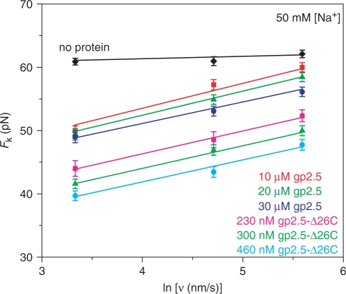
Bacteriophage T7 gene 2.5 protein (gp2.5) is a single-stranded DNA (ssDNA)-binding protein that has essential roles in DNA replication, recombination and repair. However, it differs from other ssDNA-binding proteins by its weaker binding to ssDNA and lack of cooperative ssDNA binding. By studying the rate-dependent DNA melting force in the presence of gp2.5 and its deletion mutant lacking 26 C-terminal residues, we probe the kinetics and thermodynamics of gp2.5 binding to ssDNA and double-stranded DNA (dsDNA). These force measurements allow us to determine the binding rate of both proteins to ssDNA, as well as their equilibrium association constants to dsDNA. The salt dependence of dsDNA binding parallels that of ssDNA binding. We attribute the four orders of magnitude salt-independent differences between ssDNA and dsDNA binding to nonelectrostatic interactions involved only in ssDNA binding, in contrast to T4 gene 32 protein, which achieves preferential ssDNA binding primarily through cooperative interactions. The results support a model in which dimerization interactions must be broken for DNA binding, and gp2.5 monomers search dsDNA by 1D diffusion to bind ssDNA. We also quantitatively compare the salt-dependent ssDNA- and dsDNA-binding properties of the T4 and T7 ssDNA-binding proteins for the first time.
Figures


Similar articles
-
Gp2.5, the multifunctional bacteriophage T7 single-stranded DNA binding protein.Semin Cell Dev Biol. 2019 Feb;86:92-101. doi: 10.1016/j.semcdb.2018.03.018. Epub 2018 Mar 28. Semin Cell Dev Biol. 2019. PMID: 29588157 Free PMC article. Review.
-
Interaction of bacteriophage T4 and T7 single-stranded DNA-binding proteins with DNA.Phys Biol. 2009 Jul 1;6(2):025002. doi: 10.1088/1478-3975/6/2/025002. Phys Biol. 2009. PMID: 19571366 Free PMC article.
-
Salt dependent binding of T4 gene 32 protein to single and double-stranded DNA: single molecule force spectroscopy measurements.J Mol Biol. 2005 Jun 3;349(2):317-30. doi: 10.1016/j.jmb.2005.03.065. Epub 2005 Apr 9. J Mol Biol. 2005. PMID: 15890198
-
Mechanical measurement of single-molecule binding rates: kinetics of DNA helix-destabilization by T4 gene 32 protein.J Mol Biol. 2004 Feb 27;336(4):851-70. doi: 10.1016/j.jmb.2003.12.025. J Mol Biol. 2004. PMID: 15095865
-
Biophysical characterization of DNA binding from single molecule force measurements.Phys Life Rev. 2010 Sep;7(3):299-341. doi: 10.1016/j.plrev.2010.06.001. Epub 2010 Jun 4. Phys Life Rev. 2010. PMID: 20576476 Free PMC article. Review.
Cited by
-
Gp2.5, the multifunctional bacteriophage T7 single-stranded DNA binding protein.Semin Cell Dev Biol. 2019 Feb;86:92-101. doi: 10.1016/j.semcdb.2018.03.018. Epub 2018 Mar 28. Semin Cell Dev Biol. 2019. PMID: 29588157 Free PMC article. Review.
-
Structural Basis for DNA Recognition of a Single-stranded DNA-binding Protein from Enterobacter Phage Enc34.Sci Rep. 2017 Nov 14;7(1):15529. doi: 10.1038/s41598-017-15774-y. Sci Rep. 2017. PMID: 29138440 Free PMC article.
-
Optical tweezers experiments resolve distinct modes of DNA-protein binding.Biopolymers. 2009 Apr;91(4):265-82. doi: 10.1002/bip.21123. Biopolymers. 2009. PMID: 19173290 Free PMC article. Review.
-
Single-molecule views of protein movement on single-stranded DNA.Annu Rev Biophys. 2012;41:295-319. doi: 10.1146/annurev-biophys-042910-155351. Epub 2012 Feb 23. Annu Rev Biophys. 2012. PMID: 22404684 Free PMC article. Review.
-
C-terminal phenylalanine of bacteriophage T7 single-stranded DNA-binding protein is essential for strand displacement synthesis by T7 DNA polymerase at a nick in DNA.J Biol Chem. 2009 Oct 30;284(44):30339-49. doi: 10.1074/jbc.M109.024059. Epub 2009 Sep 2. J Biol Chem. 2009. PMID: 19726688 Free PMC article.
References
-
- Chase JW, Williams KR. Single-stranded DNA binding proteins required for DNA replication. Annu. Rev. Biochem. 1986;55:103–136. - PubMed
-
- He Z-G, Rezende LF, Willcox S, Griffith JD, Richardson CC. The carboxyl-terminal domain of bacteriophage T7 single-stranded DNA-binding protein modulates DNA binding and interaction with T7 DNA polymerase. J. Biol. Chem. 2003;278:29538–29545. - PubMed
-
- He Z-G, Richardson CC. Effect of single-stranded DNA-binding proteins on the helicase and primase activities of the bacteriophage T7 gene 4 protein. J. Biol. Chem. 2004;279:22190–22197. - PubMed
-
- Kim YT, Richardson CC. Acidic carboxyl-terminal domain of gene 2.5 protein of bacteriophage T7 is essential for protein-protein interactions. J. Biol. Chem. 1994;269:5270–5278. - PubMed
-
- Kim YT, Tabor S, Churchich JE, Richardson CC. Interactions of gene 2.5 protein and DNA polymerase of bacteriophage T7. J. Biol. Chem. 1992;267:15032–15040. - PubMed
