Review
. 2008 Oct;36(17):5678-94.
doi: 10.1093/nar/gkn550. Epub 2008 Sep 4.

Gamma-H2AX in recognition and signaling of DNA double-strand breaks in the context of chromatin

Affiliations
Review

Gamma-H2AX in recognition and signaling of DNA double-strand breaks in the context of chromatin

Andrea Kinner et al. Nucleic Acids Res. 2008 Oct.

Abstract

DNA double-strand breaks (DSBs) are extremely dangerous lesions with severe consequences for cell survival and the maintenance of genomic stability. In higher eukaryotic cells, DSBs in chromatin promptly initiate the phosphorylation of the histone H2A variant, H2AX, at Serine 139 to generate gamma-H2AX. This phosphorylation event requires the activation of the phosphatidylinositol-3-OH-kinase-like family of protein kinases, DNA-PKcs, ATM, and ATR, and serves as a landing pad for the accumulation and retention of the central components of the signaling cascade initiated by DNA damage. Regions in chromatin with gamma-H2AX are conveniently detected by immunofluorescence microscopy and serve as beacons of DSBs. This has allowed the development of an assay that has proved particularly useful in the molecular analysis of the processing of DSBs. Here, we first review the role of gamma-H2AX in DNA damage response in the context of chromatin and discuss subsequently the use of this modification as a surrogate marker for mechanistic studies of DSB induction and processing. We conclude with a critical analysis of the strengths and weaknesses of the approach and present some interesting applications of the resulting methodology.


Figures
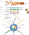
Figure 1.
Figure 1.
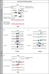
H2AX in the context of chromatin. (A) Organization of DNA in chromatin. One hundred and forty-seven base pairs of DNA (red) are wrapped around a nucleosome (yellow) consisting of eight histone proteins (two H2A/H2B dimers and two H3/H4 dimers), thus forming the 11 nm nucleosome. The histones dimerize via the histone fold motif and four histone dimers form the nucleosome core. Nucleosomes are separated by linker DNA sections of 20–80 bp in length. The DNA wraps in 1.7 turns around the nucleosome forming 142 hydrogen bonds at the DNA histone interface. The histone tails protrude from the nucleosome core and can be modified, for instance by acetylation, phosphorylation or ubiquitinylation. Further condensation of chromatin, as in the 30 nm fiber, allows a 100-fold compaction of DNA (schematic representation; the actual organization of the nucleosomes in the 30 nm fiber is still under investigation). (B) All histone proteins share the highly conserved histone fold motif (displayed in color) containing the three alpha helices involved in nucleosome core organization. Alpha helical domains outside the histone fold domain are shown in gray. The structure on the right illustrates how two histone fold domains interact for dimer formation. (C) A model of the nucleosome core particle showing DNA interactions with core histones (redrawn in a modified form from Ref. (148)).The DNA entry and exit points are localized at the H2A/H2B dimer. The H2AX C-terminus, which is 14 amino acids longer than that of H2A, is drawn here (there are no structural data available and the schematic drawing is only for demonstration purposes) in black with a red arrow marking the phosphorylation site within the SQEY motif.
Figure 2.
Figure 2.
DSB repair pathways. (A) Homologous recombination repair (HRR). After the initial sensing of the DSB by MRN and the activation of ATM, H2AX is phosphorylated, which in turn elicits a sequence of signaling events thought to ultimately cause the activation of nucleases such as Mre11 and CtIP to process the DNA ends and generate ssDNA with 3′ overhangs. ssDNA is bound by RPA, which is subsequently exchanged by Rad51 and Rad51 paralogs. This exchange is facilitated by Rad52, Rad54 and BRCA2. The Rad51-decorated DNA fiber initiates strand invasion into an intact homologous DNA molecule that leads to the formation of a Holiday junction. The DNA sequence around the DSB is copied by DNA synthesis associated with branch migration, and the process is completed by resolution of the Holiday junction. HRR is a templated repair process and is therefore error free (15–17). (B) DNA-PK-dependent nonhomologous end joining (D-NHEJ). DNA ends are recognized by Ku, which recruits, after processing by Tdp1 or PNKP, DNA-PKcs. Upon end-binding, DNA-PKcs is activated and phosphorylates itself and possibly also other proteins (like H2AX on an adjacent nucleosome). Phosphorylated DNA-PKcs is thought to be released from the DNA end, which allows the DNA ligase IV/XRCC4/XLF complex to mediate end-ligation possibly with the help of a DNA polymerase that catalyzes gap filling (19,149,150). (C) Back up pathway of nonhomologous end joining (B-NHEJ). There is evidence that cells of higher eukaryotes with defects in D-NHEJ rejoin the majority of DSBs using an alternative repair pathway that is not utilizing any of the HRR-associated activities (19). This pathway is therefore termed backup NHEJ (B-NHEJ). Although details of this pathway remain to be elucidated, there is evidence that it utilizes the PARP-1/DNA Ligase III/XRCC1 repair module known to be involved in the repair of SSB and base damages (151–155), and that its function is facilitated by the linker histone H1 (156). (D) Single strand annealing (SSA). This repair pathway shares features of HRR and NHEJ, and is best described in yeast (17). After the initial sensing of the DSBs and processing of the ends by an exonuclease, possibly the MRN complex, the generated ssDNA tails are loaded with RPA. Ends are resected until homologous regions are exposed on the two DNA strands and pairing of these regions is facilitated by Rad52. After appropriate gap filling and removal of the overhangs by the ERCC1/XPF nuclease, a ligation step restores DNA integrity. The repair pathways described in B, C and D are associated with loss (and sometimes gain) of DNA material and are by nature error prone.
Figure 3.
Figure 3.
Conserved H2AX variants. H2AX is highly conserved through evolution. Shown here is an amino acid sequence comparison between human, mouse, Xenopus and Drosophila. Identical amino acids are shown in dark boxes, and light boxes display blocks of similar amino acids. Light grey letters indicate nonsimilar amino acids, grey letters conserved and black letters weakly similar amino acids. The SQEY motif is underlined, and Serine 139, the residue that becomes phosphorylated upon induction of DSBs, is displayed in red. H2AX protein sequences were obtained from Swiss-Prot/TrEMBL and aligned using the VectorNTI software.
Figure 4.
Figure 4.
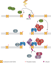
γ-H2AX is a specific and efficient coordinator of DDR signaling. Following the initial phosphorylation of H2AX by ATM, or DNA-PK, a nucleation reaction is initiated starting with the recruitment of MDC1 and continuing with that of the MRN complex to further activate ATM. This generates a feedback loop that leads to further phosphorylation of H2AX and the chromatin modifications required for the recruitment of 53BP1. The activation cascade culminates with the recruitment of RNF8 to phosphorylated MDC1 and the polyubiquitinylation of H2AX to recruit BRCA1/BARD1 (see text for details).
Figure 5.
Figure 5.
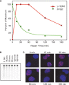
Comparison of DSB repair kinetics as measured by pulsed-field gel electrophoresis (PFGE) with the development of γ-H2AX foci. (A) Plateau-phase A549 cells were exposed to 20 Gy or 1 Gy X-rays and analyzed by PFGE (157) or γ-H2AX immunofluorescence (158), respectively, at the indicated time points. PFGE results (squares) have been normalized to the signal measured at 0 h, while the γ-H2AX results (circles) have been normalized to the maximum number of foci scored. The number of foci per cell was quantified using the Leica Q-Win software with the help of a special routine developed for foci counting. Foci were counted on 3D picture stacks generated on a Leica SP5 confocal microscope. γ-H2AX results were normalized to the maximum number of foci scored per cell—typically reached between 30 min and 1 h after IR. (B) Typical gel used to generate the PFGE results shown in A. DNA is stained with ethidium bromide. (C) Examples of γ-H2AX immunofluorescence at different times after exposure to IR (1 Gy). Cells were fixed with 2% paraformaldehyde, permeabilized with 0.2% Triton X-100 and stained with a phosphospecific primary anti-γ-H2AX antibody and an Alexa 568-labeled secondary antibody. Red shows γ-H2AX foci, blue nuclei stained with DAPI.
Figure 6.
Figure 6.
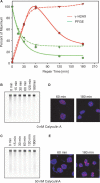
Effect of the phosphatase inhibitor Calyculin A on DSB repair kinetics and γ-H2AX foci development and decay. (A) Plateau-phase A549 cells were incubated with 0 or 50 nM Calyculin A 15 min prior to exposure to 20 or 1 Gy X-rays for PFGE or γ-H2AX immunofluorescence, respectively, and allowed to repair at 37°C for the indicated periods of time (other details of experimental design as in Figure 5). Results are shown normalized as described in Figure 5. (B, C) Typical PFGE gels used to generate the results shown in A for cells treated with 0 or 50 nM Calyculin A. DNA is stained with ethidium bromide. (D, E) γ-H2AX immunofluorescence at different times after irradiation and incubation with 0 or 50 nM Calyculin A. Other details as in Figure 5.
Figure 7.
Figure 7.
γ-H2AX foci are preferentially formed in regions of euchromatin. Elutriated G2 cells of DSB repair-deficient LIG4−/− MEFs were exposed to 16 Gy X-rays and analyzed for γ-H2AX immunofluorescence 3 h later. The picture on the left shows a DAPI-stained nucleus. Bright areas correspond to nuclear regions with increased DNA presence, thought to reflect densely packaged heterochromatin. The picture at the center shows γ-H2AX immunofluorescence obtained as described in Figure 5. The picture on the right shows an overlay of the two images with DNA displayed in blue and γ-H2AX foci in red. Note the nearly complete absence of γ-H2AX foci from heterochromatic areas, as well as from areas in the nucleus with reduced amounts of DNA.

Similar articles

Cited by

References

    1. Luger K, Mäder AW, Richmond RK, Sargent DF, Richmond S, Richmond TJ. Crystal structure of the nucleosome core particle at 2.8 Å resolution. Nature. 1997;389:251–260. - PubMed
    1. Loyola A, Almouzni G. Histone chaperones, a supporting role in the limelight. Biochim. Biophys. Acta. 2004;1677:3–11. - PubMed
    1. Cremer T, Cremer C. Chromosome territories, nuclear architecture and gene regulation in mammalian cells. Nat. Rev. Genet. 2001;2:292–301. - PubMed
    1. Misteli T. Beyond the sequence: cellular organization of genome function. Cell. 2007;128:787–800. - PubMed
    1. Tremethick DJ. Higher-order structures of chromatin: the elusive 30 nm fiber. Cell. 2007;128:651–668. - PubMed

Publication types