. 2009 Jan;150(1):324-32.
doi: 10.1210/en.2008-0988. Epub 2008 Sep 4.

Progesterone influence on neurite outgrowth involves microglia

Affiliations

Progesterone influence on neurite outgrowth involves microglia

Angela M Wong et al. Endocrinology. 2009 Jan.

Abstract

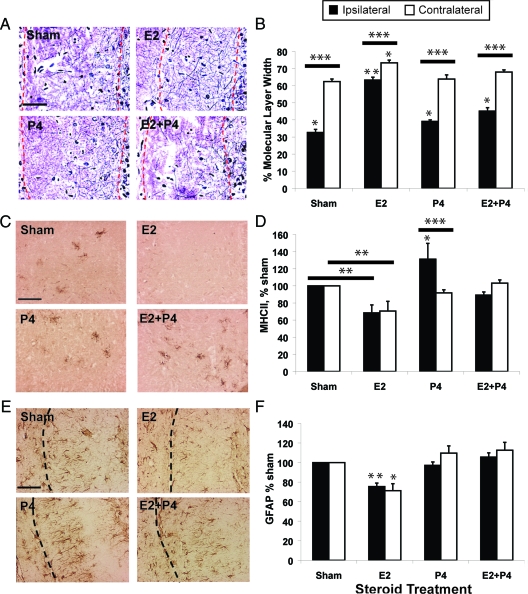
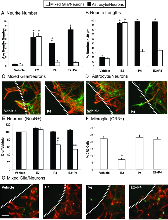
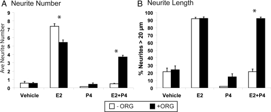
Progesterone (P4) antagonizes estradiol (E2) in synaptic remodeling in the hippocampus during the rat estrous cycle. To further understand how P4 modulates synaptic plasticity, we used entorhinal cortex lesions, which induce E2-dependent neurite sprouting in the hippocampus. In young ovariectomized rats, the E2-dependent entorhinal cortex lesion-induced sprouting was attenuated by concurrent treatment with P4 and E2. Microglial activation also showed the E2-P4 antagonism. These findings extend reports on the estrous cycle synaptic remodeling without lesions by showing the P4-E2 antagonism during simultaneous treatment with both E2 and P4. Glial mechanisms were analyzed with the wounding-in-a-dish model of cocultured glia and embryonic d-18 cortical neurons from rat. In cocultures of mixed glia (astrocytes plus 30% microglia), P4 antagonized the E2-dependent neurite outgrowth (number and length) and neuron viability in the presence of E2, as observed in vivo. However, removal of microglia (astrocyte-neuron coculture) abolished the antagonism of E2 by P4 on neuron sprouting. The P4 receptor antagonists ORG-31710 and RU-486 blocked the antagonism of P4 on E2-dependent sprouting. These findings suggest a new role for microglia in P4 antagonism of E2 in neuronal plasticity and show its dependence on progesterone receptors. These findings are also relevant to the inclusion of progestins in hormone therapy, which is controversial in relation to cognitive declines during aging and in Alzheimer's disease.


Figures

Figure 1
Figure 1
ECL in the rodent brain are a model for the perforant path degeneration in AD, which damages a major input from the cerebral cortex to the hippocampus, a key center of declarative memory. Perforant path axons arise from large neurons in layers II and III of the lateral entorhinal cortex (EC) and project to dendrites of granule cell neurons in the outer molecular layer (OML) of the dentate gyrus (DG). The hippocampal fissure is an embryonic remnant that physically separates the dentate gyrus from the dendritic field of CA1 neurons. The granule cell axons (mossy fibers) project to CA3 pyramidal neurons, which in turn send Schaffer collateral axons to CA1 pyramidal neurons. These three links comprise the trisynaptic circuit of the hippocampal memory module. Degeneration of EC neurons and perforant path axons arises early in AD, whereas the granule and CA3 neurons are relatively spared throughout AD. After unilateral ECL, compensatory sprouting (reinnervation) within the molecular layer of the dentate gyrus arises from multiple pathways; the commissural/associational axons innervate the inner molecular layer (IML), and axons from the septohippocampal pathway, from the contralateral EC, and from local interneurons sprout to innervate the outer molecular layer (OML). Inset shows Holmes axonal fiber stain.
Figure 2
Figure 2
P4 attenuates E2-induced compensatory sprouting after ipsilateral ECL (see Fig. 1). All rats were ovariectomized before ECL and killed 14 d after ECL. Controls received sham pellets without steroids. Scale bars, 100 μm. n = 8 rats per group, 3 sections per brain. Results are means ± sem. A, Axonal fibers (Holmes stained) in the ipsilateral molecular layer of the dentate gyrus (borders outlined by red dashed lines). B, Average fiber band width in molecular layers of the ipsilateral (lesioned) and contralateral (unlesioned) sides, expressed as percent total molecular layer width (granule neuron layer edge to fissure). *, P < 0.05 and **, P < 0.001 from other groups in respective hippocampal side; ***, P < 0.0001, ipsi- vs. contralateral side. C, Microglial activation in the hippocampal hilus by OX6 immunostaining (epitope for MhcII 1a peptides). D, OX6-immunostained area in ipsi- and contralateral hilus, as percentage of sham. *, P < 0.05 and **, P < 0.001 from other treatments on respective side; *** P < 0.01, ipsi- vs. contralateral side. E, Astrocyte GFAP immunostaining in the ipsilateral molecular layer of the dentate gyrus. Dashed line shows the hippocampal fissure. F, GFAP-immunostained area in ipsi- and contralateral hippocampus, as percentage of sham. *, P < 0.01 and **, P < 0.005 vs. other treatments on respective hippocampal side.
Figure 3
Figure 3
Wounding-in-a-dish model of lesions to evaluate whether the presence of microglia influences responses of neurite outgrowth in the presence of E2 and P4 in culture media. Glia were cocultured with E18 neurons for 3 d and subjected to scratch wounding, with subsequent analysis 48 h later. Green, neurite MAP-5 immunostaining; red, astrocytic GFAP immunostaining; scale bar, 100 μm; dashed line, scratch wound zone. Results show the average of three experiments ± sem. A, Numbers of neurites extended into the wound zone per 0.5 mm2 in cultures of astrocytes (black bars) or mixed glia (white bars). *, P < 0.001. B, Neurite outgrowth, expressed as percentage of neurites bigger than 20 μm (legend as for A). *, P < 0.001. C, Addition of P4 alone did not induce neurite outgrowth in mixed glia-neuron cocultures containing 30% microglia and 70% astrocytes. D, Addition of P4 alone induced neurite outgrowth in astrocyte-neuron cocultures. E, NeuN-positive neurons in 0.250 mm2 adjacent to wound zone. *, P < 0.0005, vehicle and E2 in respective coculture model; **, P < 0.0001, vehicle and E2. F, Histogram of microglia adjacent to wound zone showing percentage of cells immunoreactive for CR3/total glial cells (CR3+GFAP); *, P < 0.0001 from other treatments. G, Glia in mixed glial-neuron cocultures with steroid treatment. Immunostaining for astrocytes GFAP (red) and microglia CR3 (OX42 antibody, green).
Figure 4
Figure 4
The P4 receptor antagonist ORG (100 nm) blocked the antagonism by P4 of E2-induced neurite outgrowth in mixed glial-neuron cocultures. A, Neurite number (total); B, neurites bigger than 20 μm in the wound zone. Results are the average of three experiments ± sem. *, P < 0.0001 for −ORG vs. +ORG.

Similar articles

Cited by

References

    1. Woolley CS, McEwen BS 1993 Roles of estradiol and progesterone in regulation of hippocampal dendritic spine density during the estrous cycle in the rat. J Comp Neurol 336:293–306 - PubMed
    1. Choi JM, Romeo RD, Brake WG, Bethea CL, Rosenwaks Z, McEwen BS 2003 Estradiol increases pre- and post-synaptic proteins in the CA1 regions of the hippocampus in female rhesus macaques (Macaca mulatta). Endocrinology 144:4734–4738 - PubMed
    1. McEwen BS 2002 Estrogens effects on the brain: multiple sites and molecular mechanisms. J Appl Physiol 91:2785–2801 - PubMed
    1. Chen E, Nilsen J, Brinton RD 2006 Dose and temporal pattern of estrogen exposure determines neuroprotective outcome in hippocampal neurons: therapeutic implications. Endocrinology 147:5303–5313 - PubMed
    1. Carroll JC, Rosario ER, Chang L, Stanczyk FZ, Oddo S, LaFerla FM, Pike CJ 2007 Progesterone and estrogen regulate Alzheimer-like neuropathology in female 3xTg-AD mice. J Neurosci 27:13357–13365 - PMC - PubMed

Publication types