. 2009 Jan;27(1):230-7.
doi: 10.1634/stemcells.2008-0273.

IFATS collection: Human adipose tissue-derived stem cells induce angiogenesis and nerve sprouting following myocardial infarction, in conjunction with potent preservation of cardiac function

Affiliations

IFATS collection: Human adipose tissue-derived stem cells induce angiogenesis and nerve sprouting following myocardial infarction, in conjunction with potent preservation of cardiac function

Liying Cai et al. Stem Cells. 2009 Jan.

Abstract

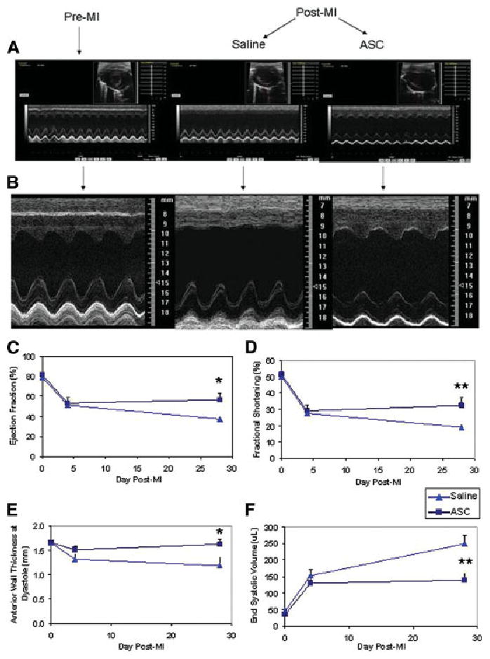
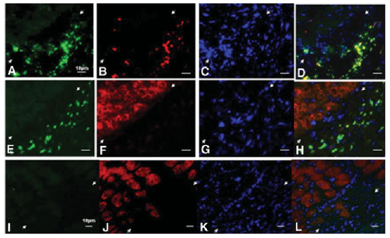
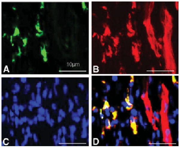
The administration of therapeutic cell types, such as stem and progenitor cells, has gained much interest for the limitation or repair of tissue damage caused by a variety of insults. However, it is still uncertain whether the morphological and functional benefits are mediated predominantly via cell differentiation or paracrine mechanisms. Here, we assessed the extent and mechanisms of adipose-derived stromal/stem cells (ASC)-dependent tissue repair in the context of acute myocardial infarction. Human ASCs in saline or saline alone was injected into the peri-infarct region in athymic rats following left anterior descending (LAD) coronary artery ligation. Cardiac function and structure were evaluated by serial echocardiography and histology. ASC-treated rats consistently exhibited better cardiac function, by all measures, than control rats 1 month following LAD occlusion. Left ventricular (LV) ejection fraction and fractional shortening were improved in the ASC group, whereas LV remodeling and dilation were limited in the ASC group compared with the saline control group. Anterior wall thinning was also attenuated by ASC treatment, and post-mortem histological analysis demonstrated reduced fibrosis in ASC-treated hearts, as well as increased peri-infarct density of both arterioles and nerve sprouts. Human ASCs were persistent at 1 month in the peri-infarct region, but they were not observed to exhibit significant cardiomyocyte differentiation. Human ASCs preserve heart function and augment local angiogenesis and cardiac nerve sprouting following myocardial infarction predominantly by the provision of beneficial trophic factors.


Conflict of interest statement

Disclosure of Potential Conflicts of Interest

B.H.J. and K.L.M. have performed contract work for Lilly.

Figures

Figure 1
Figure 1
Serial echocardiographic analysis of heart function demonstrates a functional improvement due to ASC treatment. (A): Examples of two-dimensional (2D) and M-mode echocardiographic images obtained from a normal heart before infarction (pre-MI) and then again at 28 days after MI and injections with either saline or ASCs. An evident deficit in the anterior wall motion (top of each image) was detected, which was partially ameliorated by ASC treatment. (B): Enlarged M-mode images shown in (A). (CF): Graphs of functional parameters obtained from echocardiographic images (mean ± SEM; n = 10 for each value). Parameters shown are ejection fraction as determined from 2D images (C), fractional shortening as determined from 2D images (D), anterior wall thickness as determined from M-mode images (E), and end-systolic volume as determined from 2D images (F). A complete listing of parameters is shown in Table 1. *, p < .05; **, p < .01. Abbreviations: ASC, adipose-derived stromal/stem cell; MI, myocardial infarction.
Figure 2
Figure 2
Injection of acutely infarcted hearts with ASCs attenuated the extent of infarction. Images of thin sections of a normal heart (A) or hearts at 28 days after infarction and treatment with either saline (B) or ASCs (C). Red indicates viable myocardium; blue indicates fibrosis due to infarction damage. The percentage of infarcted left ventricular area was determined by planimetry, and the average ± SEM for all hearts (n = 10) in the two groups is shown (D). The infarction area was reduced in ASC-treated hearts (26% ± 6% vs. 34% ± 6% in saline) (*, p < .05). Abbreviation: ASC, adipose-derived stromal/stem cell.
Figure 3
Figure 3
Microvessel densities within the border zone of the infarct were enhanced by treatment with ASCs. Immunohistochemical analysis of thin sections from each heart (n = 10 for each group) was performed with antibodies to smooth muscle α-actin (SMA). The density of smooth muscle SMA+ arterioles was determined in the remote posterior regions, as well as border and infarct zones, as described in Materials and Methods. The number of microvessels per area was greater in ASC-treated hearts than in the saline control group (*, p < .05). Abbreviation: ASC, adipose-derived stromal/stem cell.
Figure 4
Figure 4
Treatment with ASCs promotes enhanced nerve sprouting in viable peri-infarction regions of the heart. (A): Representative photomicrograph of growth-associated protein-43 (GAP43)+ nerve sprouts (red arrow) in the left ventricular region 1 month after infarction and treatment with ASCs. (B): ASCs induced more GAP43+ nerve sprouts in the border zones of the infarct (464 ± 43 μm2/mm2) compared with the control group (342 ± 20 μm2/mm2) (*, p < .05). There was no significant difference between the two groups in the posterior myocardial wall nerve sprouting. Abbreviation: ASC, adipose-derived stromal/stem cell.
Figure 5
Figure 5
Viable adipose tissue-derived stromal/stem cells (ASCs) stably engraft in the regions adjacent to, but distinct from, viable cardiomyocytes. (A): Immunofluorescent detection of donor ASCs was accomplished by probing for human leukocyte antigen (HLA)-ABC (green). The ASCs were located within the infarct region (white arrows in all panels demarcate the border zone). (B): These ASCs were viable and actively proliferating as evidenced by the expression of the Ki67 marker (red). (C): Visualization of nuclei with 4′,6-diamidino-2-phenylindole dihydrochloride (DAPI) stain. (D): Merged images shown in (A–C) demonstrate colocalization of Ki67 and HLA-ABC staining (yellow). (E–H): Clusters of ASCs were often observed in proximity to live myocardium within peri-infarct regions. (E): Immunofluorescent images of HLA-ABC-expressing ASCs (green). (F, G): Viable α-sarcomeric actin+ cardiomyocytes (red staining in (F)) and DAPI-stained nuclei (blue staining in (G)). (H): The merged images (E–G) provides evidence that ASCs reside in juxtaposition with cardiomyocytes and are not present within viable myocardium. (I): Probing similar sections with an isotype control antibody demonstrated the specificity of the HLA-ABC antibody for human-derived ASCs. (J, K): Costaining of the section shown in (I) with an antibody to α-sarcomeric actin and DAPI nuclear stain. (L): Merged images (IK). Scale bars = 10 μm.
Figure 6
Figure 6
Long-term engraftment of adipose tissue-derived stromal/ stem cells (ASCs) occurs in proximity to vascular structures. (A–D): Immunofluorescent micrographs of a thin section from an infarcted rat heart at 1 month after treatment with ASCs. (A): Detection of human leukocyte antigen (HLA)-ABC+ ASCs (green). (B, C): The same section probed with antibodies to smooth muscle α-actin (SMA) (red) (B) and 4′,6-diamidino-2-phenylindole dihydrochloride nuclear stain (blue) (C). (D): Merged images (A–C) show the coexpression of HLA-ABC and SMA antigens by the ASCs. A branching vessel is evident in the right portion of the image. Scale bars = 10 μm.

References

    1. Abdel-Latif A, Bolli R, Tleyjeh IM, et al. Adult bone marrow-derived cells for cardiac repair: A systematic review and meta-analysis. Arch Intern Med. 2007;167:989–997. - PubMed
    1. Erbs S, Linke A, Schachinger V, et al. Restoration of microvascular function in the infarct-related artery by intracoronary transplantation of bone marrow progenitor cells in patients with acute myocardial infarction: The Doppler Substudy of the Reinfusion of Enriched Progenitor Cells and Infarct Remodeling in Acute Myocardial Infarction (REPAIR-AMI) trial. Circulation. 2007;116:366–374. - PubMed
    1. Lipinski MJ, Biondi-Zoccai GG, Abbate A, et al. Impact of intracoronary cell therapy on left ventricular function in the setting of acute myocardial infarction: A collaborative systematic review and meta-analysis of controlled clinical trials. J Am Coll Cardiol. 2007;50:1761–1767. - PubMed
    1. Cao Y, Sun Z, Liao L, et al. Human adipose tissue-derived stem cells differentiate into endothelial cells in vitro and improve postnatal neovascularization in vivo. Biochem Biophys Res Commun. 2005;332:370–379. - PubMed
    1. Planat-Benard V, Silvestre JS, Cousin B, et al. Plasticity of human adipose lineage cells toward endothelial cells: Physiological and therapeutic perspectives. Circulation. 2004;109:656–663. - PubMed

Publication types

MeSH terms