. 2008 Oct;173(4):1085-99.
doi: 10.2353/ajpath.2008.080382. Epub 2008 Sep 4.

Smad-independent transforming growth factor-beta regulation of early growth response-1 and sustained expression in fibrosis: implications for scleroderma

Affiliations

Smad-independent transforming growth factor-beta regulation of early growth response-1 and sustained expression in fibrosis: implications for scleroderma

Swati Bhattacharyya et al. Am J Pathol. 2008 Oct.

Abstract

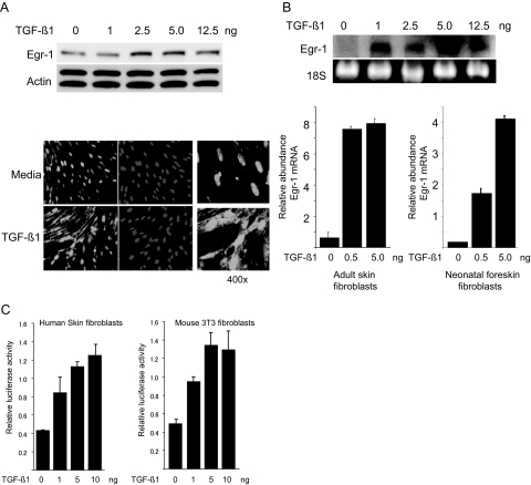
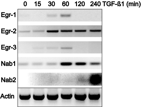
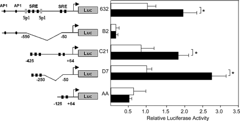
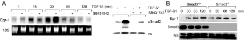
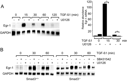
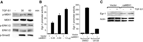
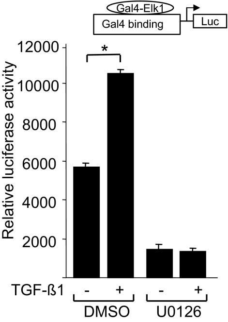
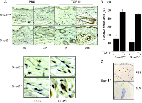
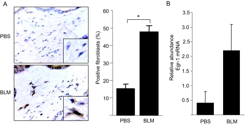
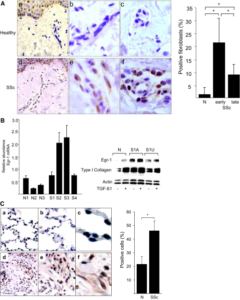
Transforming growth factor-beta (TGF-beta) plays a key role in scleroderma pathogenesis. The transcription factor early growth response-1 (Egr-1) mediates the stimulation of collagen transcription elicited by TGF-beta and is necessary for the development of pulmonary fibrosis in mice. Here, we report that TGF-beta causes a time- and dose-dependent increase in Egr-1 protein and mRNA levels and enhanced transcription of the Egr-1 gene via serum response elements in normal fibroblasts. The ability of TGF-beta to stimulate Egr-1 was preserved in Smad3-null mice and in explanted Smad3-null fibroblasts. The response was blocked by a specific mitogen-activated protein kinase kinase 1 (MEK1) inhibitor but not by an ALK5 kinase inhibitor. Furthermore, MEK1 was phosphorylated by TGF-beta, which was sufficient to drive Egr-1 transactivation. Stimulation by TGF-beta enhanced the transcriptional activity of Elk-1 via the MEK-extracellular signal-regulated kinase 1/2 pathway. Bleomycin-induced scleroderma in the mouse was accompanied by increased Egr-1 accumulation in lesional fibroblasts. Furthermore, biopsies of lesional skin and lung from patients with scleroderma showed increased Egr-1 levels, which were highest in early diffuse disease. Moreover, both Egr-1 mRNA and protein were elevated in explanted scleroderma skin fibroblasts in vitro. Together, these findings define a Smad-independent TGF-beta signal transduction mechanism that underlies the stimulation of Egr-1, demonstrate for the first time sustained Egr-1 up-regulation in fibrotic lesions and suggests that Egr-1 has a role in the induction and progression of fibrosis.


Figures

Figure 1
Figure 1
TGF-β stimulates Egr-1 expression in fibroblasts. Serum-starved neonatal human foreskin fibroblasts (A–C) or adult skin fibroblasts (B) or mouse NIH3T3 fibroblasts (C) at confluence were incubated with indicated concentrations of TGF-β1. A: After 120 minutes incubation, whole-cell lysates were prepared and subjected to Western analysis (top). Representative immunoblots are shown. Fibroblasts incubated with 1 ng/ml TGF-β for 120 minutes were immunostained with antibodies to Egr-1 or with 4,6-diamidino-2-phenylindone and viewed by laser scanning confocal microscopy (bottom). Representative images are shown. Magnification: ×100 (left and middle panels); ×400 (right panels). B: After 30-minute incubation, total RNA was isolated and examined by Northern analysis (top) or by real-time quantitative PCR (bottom; bars indicate means ± SD from triplicate determinations). Values are the mean and SD and normalized with Actin. Representative results are shown. C: Fibroblasts were transiently transfected with Egr-1 promoter-luc constructs. After incubation of the cultures with increasing concentrations of TGF-β1 for 24 hours, cell lysates were assayed for their luciferase activities. The results are the means ± SD of triplicate determinations.
Figure 2
Figure 2
TGF-β regulation of the expression of Egr moieties. Confluent foreskin fibroblasts were incubated with TGF-β1 (12.5 ng) for indicated periods. Total RNA was isolated and subjected to RT-PCR analysis. The results are representative of at least two independent experiments.
Figure 3
Figure 3
Delineation of TGF-β response region of the Egr-1 gene promoter. NIH3T3 fibroblasts transiently transfected with indicated 5′ deletion or truncation mutants of the mouse Egr-1 gene promoter linked to the luciferase reporter were incubated with TGF-β1 for 24 hours. Cell lysates were assayed for their luciferase activities. The results, normalized with Renilla luciferase activities, are the means ± SD of triplicate determinations from at least two independent experiments. □, untreated fibroblasts; ▪, TGF-β-treated fibroblasts. *P < 0.005.
Figure 4
Figure 4
Smad3-independent TGF-β stimulation of Egr-1. A: Confluent serum-starved foreskin fibroblasts were pretreated with 10 μmol/L SB431542 for 30 minutes, followed by TGF-β1 for indicated periods. At the end of the incubation, total RNA was isolated and examined by Northern analysis. The results are representative of two independent experiments. B: Time-dependent Egr-1 stimulation in mouse skin fibroblasts. Confluent serum-starved fibroblasts explanted from Smad3-null mice and wild-type littermates in parallel were incubated with TGF-β1 for indicated periods. Whole-cell lysates were examined by Western analysis. NS, nonspecific band. Representative immunoblots are shown.
Figure 5
Figure 5
MEK1-dependent stimulation of Egr-1 mRNA. Confluent serum-starved human (A) or mouse (B) skin fibroblasts were preincubated with protein kinase inhibitors (10 μmol/L) for 30 minutes, followed by TGF-β1 for indicated periods. A: Cultures were harvested, and total RNA was subjected to Northern analysis (left) or real-time quantitative PCR analysis (right). For real-time PCR, values are the mean and SD and normalized with Actin. *P < 0.05. B: RNA from Smad3-null dermal fibroblasts or wild-type control fibroblasts was subjected to Northern analysis. Representative autoradiograms are shown.
Figure 6
Figure 6
MEK1 is activated by TGF-β and induces Egr-1. A: Foreskin fibroblasts were incubated with TGF-β for indicated periods and whole-cell lysates were subjected to Western analysis. Representative autoradiograms are shown. B: NIH3T3 fibroblasts were cotransfected with indicated concentrations of constitutively active MEK1 (caMEK1) or empty vector along with Egr-1-luc (left). After incubation for 24 hours, cultures were harvested, and cell lysates were assayed for their luciferase activities. Transfected NIH3T3 fibroblasts were incubated in media containing U0126 for 24 hours (right). Results, normalized with Renilla luciferase, are expressed as means ± SD of triplicate determinations from two independent experiments. C: Whole-cell lysates prepared from transfected NIH3T3 fibroblasts incubated with TGF-β for 24 h were examined by Western analysis. Representative immunoblots are shown.
Figure 7
Figure 7
TGF-β stimulates Elk-1-dependent transcription. Subconfluent NIH3T3 fibroblasts were cotransfected with the expression vector pGal4-dbd-Elk-1 along with the reporter construct pGal4-DNA-luc. Transfected fibroblasts were preincubated in the presence or absence of 10 μmol/L U0126 for 30 minutes before incubation with TGF-β1 for 24 hours. Cell lysates were assayed for their luciferase activities. The results, shown as the means ± SD of triplicate determinations, are representative of three independent experiments. *P < 0.01.
Figure 8
Figure 8
TGF-β induces Egr-1 expression in vivo. Six-week-old female Smad3-null mice and wild-type littermate mice received a single s.c. injection of 250 ng TGF-β1 or PBS. Lesional skin was harvested 1 or 24 hours later and examined by immunohistochemistry using antibodies against Egr-1. A: Representative photomicrographs; bottom panels, higher magnification. B: The proportion of Egr-1-positive fibroblasts in the lesional dermis (at 24 hours) was determined. The results represent the means ± SE. C: Negative control. Skin tissue from Egr-1-null mice was processed in parallel.
Figure 9
Figure 9
Bleomycin-induced skin fibrosis associated with Egr-1 expression in vivo. Six-week-old female BALB/C mice received daily s.c. injections of bleomycin (BLM) or PBS. A: At day 21, lesional skin was harvested. Immunohistochemistry using antibodies against Egr-1. Hematoxylin counterstain. Representative images are shown. Magnification: ×400; ×1000 (inset, left). The proportion of immunopositive fibroblasts in the lesional dermis was determined (right). Results represent means ± SD. B: Total RNA from lesional skin was analyzed by real-time quantitative PCR. Results are the means ± SD of duplicate determinations from three mice/group. *P < 0.005.
Figure 10
Figure 10
Elevated Egr-1 accumulation in SSc. A: Lesional skin tissue from patients with SSc (n = 23) and the healthy controls (n = 5) were processed for immunohistochemistry with antibodies against Egr-1. Representative images (left). a and d: Dermis of healthy controls and SSc, respectively (original magnification, ×400). b and e: Fibroblasts in the dermis of healthy controls and SSc tissue, respectively(original magnification, ×1000). c and f: Perivascular and vascular staining of healthy controls and SSc patients, respectively. The proportion of Egr-1-positive fibroblastic cells was calculated (right). B: SSc dermal fibroblasts explanted from lesional skin (S1–4) and control fibroblasts from healthy individuals (N1–3) were harvested, and total RNA was subjected to quantitative PCR analysis (left). The results, expressed relative to the levels of actin mRNA, are the means of triplicate determinations, and representative of two independent experiments. Whole-cell lysates from confluent fibroblasts explanted from affected (A) and unaffected (U) skin from a patient with SSc were incubated with or without TGF-β1 and subjected to Western analysis (right). Results with a representative SSc cell line (S1) are shown. Similar results were obtained with five additional SSc fibroblast lines. C: Lung tissues from patients with SSc-associated pulmonary fibrosis undergoing lung transplantation surgery (n = 4) and normal donor lungs (n = 3) were examined by immunohistochemistry as above. a–c: Normal lungs. d–f: SSc-associated pulmonary fibrosis. Magnification: ×100 (a and d); ×400 (b and e); ×1000 (c and f). Brown staining indicates Egr-1-positive cells. Nuclei are counterstained with hematoxylin (blue). The proportion of Egr-1-positive cells was determined, and results are shown as means ± SD (right). *P < 0.001.

References

    1. Jimenez SA, Derk CT. Following the molecular pathways toward an understanding of the pathogenesis of systemic sclerosis. Ann Intern Med. 2004;140:37–45. - PubMed
    1. Varga J, Abraham D. Systemic sclerosis: a prototypic multisystem fibrotic disorder. J Clin Invest. 2007;117:557–567. - PMC - PubMed
    1. Mauviel A. Transforming growth factor-beta: a key mediator of fibrosis. Methods Mol Med. 2005;117:69–80. - PubMed
    1. Massagué J, Seoane J, Wotton D. Smad transcription factors (review). Genes Dev. 2005;19:2783–2810. - PubMed
    1. Moustakas A, Heldin CH. Non-Smad TGF-beta signals (review). J Cell Sci. 2005;118:3573–3584. - PubMed

Publication types

MeSH terms