. 2008 Oct 3;322(5898):96-101.
doi: 10.1126/science.1164685. Epub 2008 Sep 4.

Internally generated reactivation of single neurons in human hippocampus during free recall

Affiliations

Internally generated reactivation of single neurons in human hippocampus during free recall

Hagar Gelbard-Sagiv et al. Science. .

Abstract

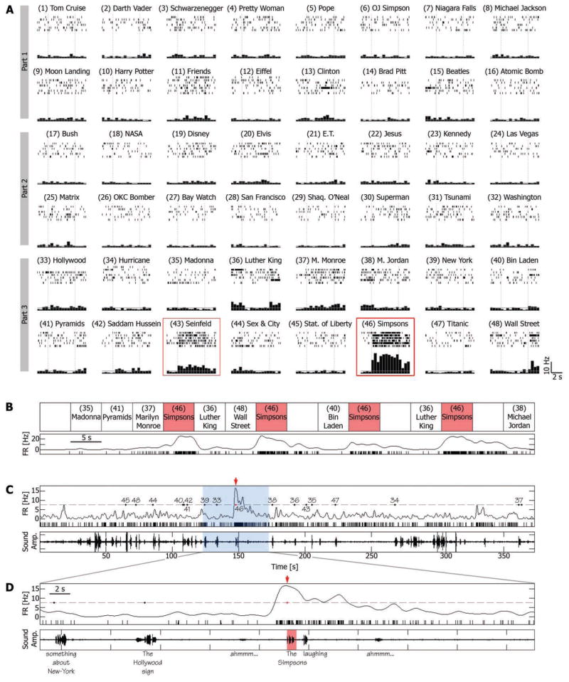
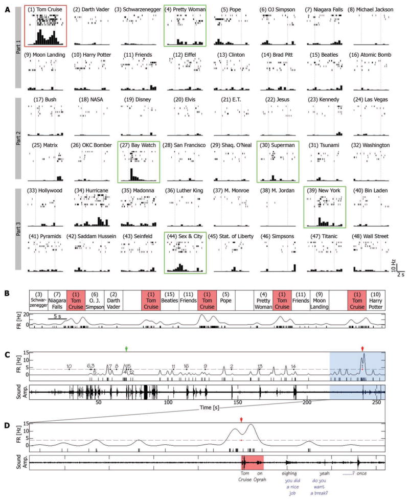
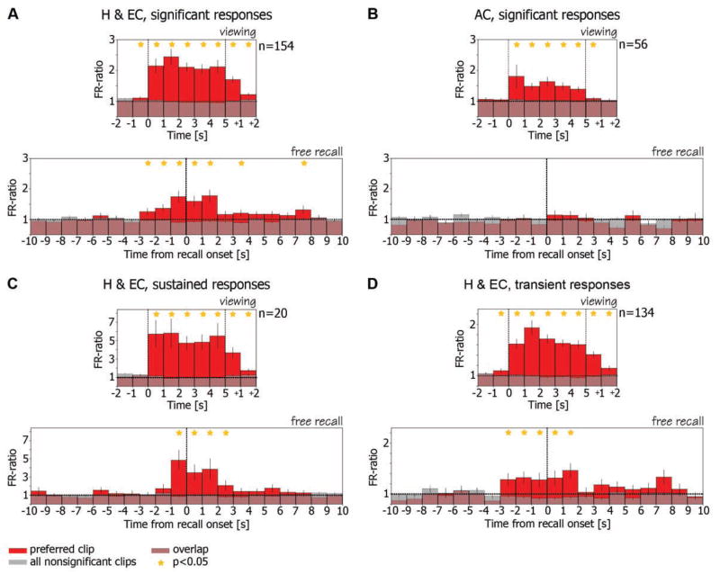
The emergence of memory, a trace of things past, into human consciousness is one of the greatest mysteries of the human mind. Whereas the neuronal basis of recognition memory can be probed experimentally in human and nonhuman primates, the study of free recall requires that the mind declare the occurrence of a recalled memory (an event intrinsic to the organism and invisible to an observer). Here, we report the activity of single neurons in the human hippocampus and surrounding areas when subjects first view cinematic episodes consisting of audiovisual sequences and again later when they freely recall these episodes. A subset of these neurons exhibited selective firing, which often persisted throughout and following specific episodes for as long as 12 seconds. Verbal reports of memories of these specific episodes at the time of free recall were preceded by selective reactivation of the same hippocampal and entorhinal cortex neurons. We suggest that this reactivation is an internally generated neuronal correlate for the subjective experience of spontaneous emergence of human recollection.


Figures

Fig. 1
Fig. 1
A single-unit in the right entorhinal cortex was activated during viewing and recall of an episode from the TV series The Simpsons. (A) Cell responses to a selection of 48 different episodes (movie clips) presented to the patient in three different viewing sessions (parts 1 to 3). For each clip, the corresponding raster plots (six trials, order of trials is from top to bottom) and post–stimulus time histogram (500-ms bins) are given. Vertical dashed lines indicate clip onset and offset (5 s apart); 5-s blank periods were presented occasionally within groups of successive clips and were used to calculate the baseline firing rate, denoted by a gray horizontal line. Red boxes indicate sustained responses. (B) Trial-by-trial response of the neuron. Order of clips is for the purpose of illustration; more intervening clips separated successive Simpsons clips in the actual experiment. Spike raster plot and instantaneous firing rate (spike train convolved with a Gaussian of the full width at half maximum of 1200 ms) are displayed together. (C) Free-recall session that followed the third viewing session (part 3). (Bottom) Sound amplitude of patient voice; (top) a spike raster plot and instantaneous firing rate; gray dashed line denotes the average firing rate during the recall session + 3 SD; numbered dots denote onset time of verbal report of recall events, corresponding to clip numbers in (A). Note the distinct elevation of firing rate just before the patient reported the recall of the Simpsons clip (red arrow). (D) A 50-s window around the Simpsons recall event [blue area in (C)]. Patient’s words are below the bottom panel. Note that the cell’s firing rate rose significantly above baseline 1500 ms before onset of verbal report of the Simpsons clip and returned to baseline after more than 10 s.
Fig. 2
Fig. 2
A single-unit in the left anterior hippocampus is activated during viewing and recall of an episode (conventions as in Fig. 1). (A and B) Note the sustained elevation of firing rate during the episode depicting actor Tom Cruise during an interview on the Oprah Winfrey Show (red box). Note also the transient responses to various clips (green boxes). (C and D) Free-recall session that followed the first viewing session (part 1). Note that the burst of spikes that accompanied the recall of the Tom Cruise clip began 1500 ms before onset of verbal report (“Tom Cruise … on Oprah”). Blue words indicate experimenter’s speech.
Fig. 3
Fig. 3
Average FR ratio histograms during viewing and during free recall. (A) (Top) Ratio of firing rate during viewing of clips to baseline firing rate is averaged across all responsive hippocampal and entorhinal cortex cells (n = 154). Vertical dashed lines denote clip onset and offset. Cells increased their firing rate significantly above baseline during and following viewing of their preferred clip (15) (red bars). Note small elevation before clip onset, probably attributable to anticipatory effect. These cells remained at baseline firing rate (FR ratio = 1) during viewing of other clips (15) (gray bars). (Bottom) FR ratio during recall events is averaged across the same cells from the top panel. Traces were aligned on the onset time of the verbal report of recall (zero time, vertical dashed line). Note that the same cells increased their firing rate significantly above baseline in the 3 s before onset of verbal report of their preferred clips (red bars) and maintained this elevated firing rate in the ensuing 2 s. However, these cells remained at baseline during recall of clips that did not elicit significant responses during viewing (gray bars). Stars denote statistical significance of P < 0.05 (t test) (15). (B) Same as (A) but for cells from anterior cingulate (n = 56). Note that, in contrast to hippocampal and entorhinal cortex cells, although these cells exhibited selectivity during viewing (top), this selectivity was not maintained during free recall (bottom). (C and D) are the same as (A) but for sustained and transient responses separately.

Comment in

References

    1. Scoville WB, Milner B. J Neurol Neurosurg Psychiatry. 1957;20:11. - PMC - PubMed
    1. Squire LR, Stark CE, Clark RE. Annu Rev Neurosci. 2004;27:279. - PubMed
    1. Eichenbaum H. Neuron. 2004;44:109. - PubMed
    1. Moscovitch M, Nadel L, Winocur G, Gilboa A, Rosenbaum RS. Curr Opin Neurobiol. 2006;16:179. - PubMed
    1. Fried I, MacDonald KA, Wilson CL. Neuron. 1997;18:753. - PubMed

Publication types