. 2008 Sep 26;321(5897):1801-6.
doi: 10.1126/science.1164368. Epub 2008 Sep 4.

Core signaling pathways in human pancreatic cancers revealed by global genomic analyses

Affiliations

Core signaling pathways in human pancreatic cancers revealed by global genomic analyses

Siân Jones et al. Science. .

Abstract

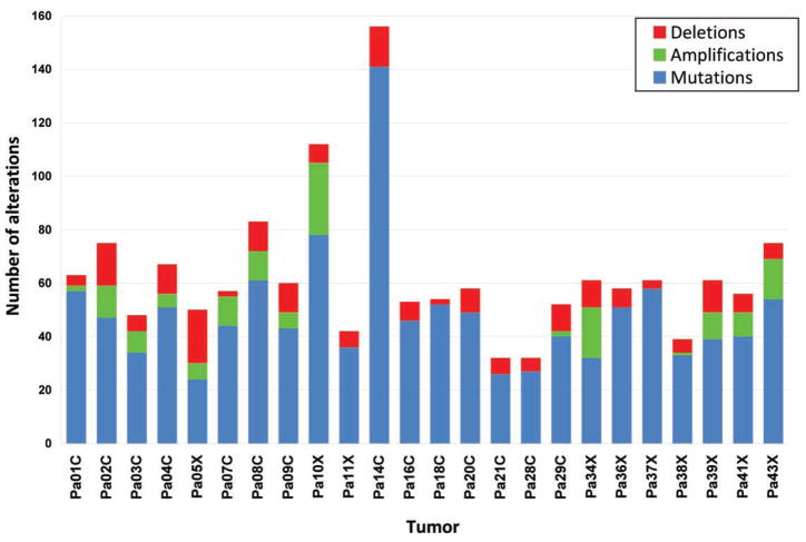
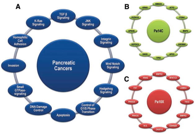
There are currently few therapeutic options for patients with pancreatic cancer, and new insights into the pathogenesis of this lethal disease are urgently needed. Toward this end, we performed a comprehensive genetic analysis of 24 pancreatic cancers. We first determined the sequences of 23,219 transcripts, representing 20,661 protein-coding genes, in these samples. Then, we searched for homozygous deletions and amplifications in the tumor DNA by using microarrays containing probes for approximately 10(6) single-nucleotide polymorphisms. We found that pancreatic cancers contain an average of 63 genetic alterations, the majority of which are point mutations. These alterations defined a core set of 12 cellular signaling pathways and processes that were each genetically altered in 67 to 100% of the tumors. Analysis of these tumors' transcriptomes with next-generation sequencing-by-synthesis technologies provided independent evidence for the importance of these pathways and processes. Our data indicate that genetically altered core pathways and regulatory processes only become evident once the coding regions of the genome are analyzed in depth. Dysregulation of these core pathways and processes through mutation can explain the major features of pancreatic tumorigenesis.


Figures

Fig. 1
Fig. 1
Examples of structural models of mutations. (A). The x-ray crystal structure of the C2 domain of protein kinase C γ (PKCG) [Protein Data Bank identification number (PDBID) 2UZP]. R252 (41) is shown as yellow space-fills; Ca2+ ions are shown as red spheres. The ligands 1,2-ethanediol and pyridoxal-5-phosphate are shown in white and purple ball-and-stick representations, respectively. The R252→H252 (R252H) mutation could reduce the membrane binding of the C2 domain of PRKCG and thereby affect function. (B) The nuclear magnetic resonance solution structure of the three tandem repeats of zf-C2H2 domains from human Kruppel-like factor 5 (KLF5) (PDBID 2EBT). H389 is shown as yellow space-fills; Zn2+ ions are shown as cyan spheres. The residues comprising the C2H2 group that coordinate the nearby Zn2+ ion are shown as ball-and-stick representations, H393 and H397 are shown in green and white, whereas C380 and C375 are shown in orange and red. The mutation at position 389 (H389N) may disrupt the structure of the zinc finger or nearby zinc coordination site. (C) The x-ray crystal structure of the heterotrimer of SMAD3 (two subunits shown as blue ribbons) and SMAD4 (one subunit shown as pink ribbons) (PDBID 1U7F). The residues corresponding to two of the mutant positions (F260S and S422F, shown as red and yellow space-fills, respectively, in chain A) are located at interfaces and could perturb Smad3-Smad3 or Smad3-Smad4 interactions. In chain B, F260 is shown as cyan space-fills and S422 as green space-fills. (D) The x-ray crystal structure of the extracellular domain of human DPP6 as a homodimer (PDBID 1XFD). Two of the mutated residues found in this study, T409I (shown as red space-fills) and D475N (shown in yellow space-fills) are in spatial proximity and are close to one of the glycosylation sites, N471 (shown as white space-fills). These mutations fall in the β-propeller domain of the protein (residues 142 to 322 and 351 to 581) thought to be involved in protein-protein interactions. The A778T mutation (shown as blue space-fills) falls in the */β hydrolase domain (residues 127 to 142 and 581 to 849) and is close to the homodimer region of the protein and could perturb the homodimer association. Carbohydrates with glycosylation sites are shown in stick representation. Images created with UCSF Chimera version 1.2422 for Linux (42).
Fig. 2
Fig. 2
Number of genetic alterations detected through sequencing and copy number analyses in each of the 24 cancers.
Fig. 3
Fig. 3
Signaling pathways and processes. (A) The 12 pathways and processes whose component genes were genetically altered in most pancreatic cancers. (B and C) Two pancreatic cancers (Pa14C and Pa10X) and the specific genes that are mutated in them. The positions around the circles in (B) and (C) correspond to the pathways and processes in (A). Several pathway components overlapped, as illustrated by the BMPR2 mutation that presumably disrupted both the SMAD4 and Hedgehog signaling pathways in Pa10X. Additionally, not all 12 processes and pathways were altered in every pancreatic cancer, as exemplified by the fact that no mutations known to affect DNA damage control were observed in Pa10X. N.O. indicates not observed.

References

    1. Parkin DM, Bray FI, Devesa SS. Eur J Cancer. 2001;37(suppl):S4. - PubMed
    1. Jemal A, et al. CA Cancer J Clin. 2008;58:71. - PubMed
    1. Koorstra JB, Hustinx SR, Offerhaus GJ, Maitra A. Pancreatology. 2008;8:110. - PMC - PubMed
    1. Efthimiou E, Crnogorac-Jurcevic T, Lemoine NR. Pancreatology. 2001;1:571. - PubMed
    1. Mimeault M, Brand RE, Sasson AA, Batra SK. Pancreas. 2005;31:301. - PubMed

Publication types

MeSH terms