. 2008 Sep 5;321(5894):1322-7.
doi: 10.1126/science.1159775.

Internally generated cell assembly sequences in the rat hippocampus

Affiliations

Internally generated cell assembly sequences in the rat hippocampus

Eva Pastalkova et al. Science. .

Abstract

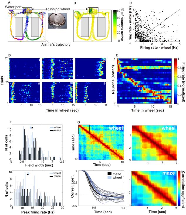
A long-standing conjecture in neuroscience is that aspects of cognition depend on the brain's ability to self-generate sequential neuronal activity. We found that reliably and continually changing cell assemblies in the rat hippocampus appeared not only during spatial navigation but also in the absence of changing environmental or body-derived inputs. During the delay period of a memory task, each moment in time was characterized by the activity of a particular assembly of neurons. Identical initial conditions triggered a similar assembly sequence, whereas different conditions gave rise to different sequences, thereby predicting behavioral choices, including errors. Such sequences were not formed in control (nonmemory) tasks. We hypothesize that neuronal representations, evolved for encoding distance in spatial navigation, also support episodic recall and the planning of action sequences.


Figures

Fig. 1
Fig. 1
“Episode fields” in the wheel and place fields in the maze are similar. (A) Color-coded spikes (dots) of simultaneously recorded hippocampal CA1 pyramidal neurons. The rat was required to run in the wheel facing to the left during the delay between the runs in the maze. (B) Percent of neurons firing >0.2 Hz within each pixel. Note the highest percentage of active neurons in the wheel. (C) Relationship between firing rate of neurons in the wheel and the maze (rs= − 0.3, p < 0,0001, 681 neurons, 3 rats, 17 sessions). (D) Normalized firing rate of six simultaneously recorded neurons during wheel running (each line shows the color-coded activity on single trials turning to the left arm). Note the transient increase of firing rate (“episode fields”) at specific segments of the run. (E) Normalized firing rate of 30 simultaneously recorded neurons during wheel running, ordered by the latency of their peak firing rate. (F) Width (top) and peak firing rate (bottom) of episode and place fields (Nwheel= 135, Nmaze=162). Arrows: medians. G. Population vector cross-correlation matrix (S.O.M.). The width of the diagonal stripe indicates the rate at which neuronal assemblies transition. Lower left: decay of population vector correlation during wheel running and maze traversal. Thin lines: individual sessions; thick lines: group means.
Fig. 2
Fig. 2
Episode neurons in the wheel display theta phase precession and ‘temporal compression”. (A) Top, unfiltered (gray) and filtered (4-10 Hz; black) traces of LFP and phase advancement of action potentials (dots). Below, activity of 6 example neurons from the same session. Each dot is an action potential, displayed as a function of theta phase and time from the beginning of wheel running from all trials. One and a half theta cycles are shown (y axis). Red line, smoothed firing rate. (B) Power spectra of spike trains generated during wheel running (n = 283 pyramidal neurons) and the simultaneously recorded LFP. Note faster oscillation of neurons relative to LFP. (C) Slope of theta phase precession within episode fields in the wheel and within place fields in the maze. (D) Relationship between phase precession slope and episode length (left, r = 0.46, p < 0.0001) and episode field width (right, r = 0.52, p < 0.0001), respectively. (E) Temporal “compression” of spikes sequences. Correlation of the distance between the peaks of episode fields of neuron pairs in the wheel with the temporal offset of the pair's cross-correlogram peak. Each dot represents a neuron pair (n = 105 eligible pairs; 3 rats; r = 0.59; p < 0.0001).
Fig. 3
Fig. 3
Firing patterns during wheel running depend on the context of the task. (A) Top, Activity of representative single neurons (color coded) during wheel running in control tasks 1 and 2 (compare with Fig. 1D). Bottom, unit discharges (dots) from all trials within a session as a function of theta phase, plotted against time from the beginning of a wheel run. Gray line, smoothed mean firing rate. Note relatively steady firing rates and steady theta phase in both control tasks. (B) Cross-correlation matrices in three different tasks (memory and control 2 are from the same rat). In the memory task, trials with the same future choices (L-trialsn vs L-trialn+1 and R-trialsn vs R-trialn+1) were cross-correlated, whereas in control tasks trialsn and trialsn+1 were cross-correlated. Only pixel values significantly different from chance are shown (Spearman rank correlation, p < 0.01). (C) Population vector correlation coefficient values in the memory task (n = 17 sessions) and control tasks (n = 8 sessions). Mean ± SD. (D) Power spectrum of spike trains of an episode neuron (unit) and simultaneously recorded LFP during wheel running in the memory task (30). Note the higher frequency of unit firing oscillation, relative to the frequency of LFP. (E) Difference between unit and LFP oscillation frequency in the memory (left) and control (right) tasks. Each line is a color-coded normalized cross-correlogram between power spectrum of a pyramidal neuron and simultaneously recorded LFP. A shift of the maximal correlation values to the right indicates that unit theta oscillation is faster than LFP theta oscillation (black dots, maxima of the cross-correlograms; white line, sum of all neurons). Note significant frequency shift in the memory task (0.44 ± 0.6Hz), and lack of frequency shift in control tasks (combined control 1 and 2: 0.07 ± 0.3Hz) (F) Ratio of spikes in the center and tail of temporal auto-correlograms (S.O.M.). High values indicate compact episode fields, low values indicate spikes scattered throughout the time of wheel running (memory task, n=287 neurons; control tasks, n = 85 neurons p < 0.0001; ranksum test). Arrows, medians.
Fig. 4
Fig. 4
Cell assembly activity in the wheel predicts future choice of the rat in the maze. (A) Examples of 3 neurons which strongly differentiated between wheel running trials preceding right and left choices (see also fig. S7 and movie 1). (B) Normalized firing rate profiles of neurons during wheel running and in the stem of the maze, ordered by the latency of their peak firing rates during left trials (each line is a single cell; cells are combined from all sessions). White line, time gap between the end of wheel running and the initiation of maze stem traversal. Middle panel: Normalized firing rates of the same neurons during right trials. Right panel: Time periods of significant differences (p < 0.05) in firing rates between left and right trials for respective neurons (red line: R > L, blue line: L > R). Grey line, number of neurons discriminating between left and right trials as a function of wheel running time.
Fig. 5
Fig. 5
Cell assembly activity in the wheel predict behavioral errors in the maze. (A) Two example neurons from a session with 7 left error trials (err). Correct trials are separated to left and right turn trials. (B) Normalized firing rates of 43 neurons simultaneously recorded during wheel running, ordered by the latency of peak firing rates during correct left trials (left). Right, firing sequence of the same neurons on correct right trials. (C) Firing sequence of neurons in a single error (left) trial. Neuronal order is the same as in B. Note that the firing sequence during the error trial is similar to that of the correct left trials. White number: correlation coefficient between correct and error trial sequences (see also fig. S13). (D) Percent of correctly predicted errors from the neuronal population activity.

Comment in

References

    1. O'Keefe J, Dostrovsky J. Brain Res. 1971;34:171–175. - PubMed
    1. O'Keefe J, Nadel L. The hippocampus as a cognitive map. Clarendon Press; Oxford, UK: 1978.
    1. Huxter J, Burgess N, O'Keefe J. Nature. 2003;425:828–832. - PMC - PubMed
    1. McNaughton BL, Barnes CA, O'Keefe J. Exp. Brain Res. 1983;52:41–49. - PubMed
    1. O'Keefe J, Burgess N. Nature. 1996;381:425–8. - PubMed

Publication types