. 2008 Nov 15;112(10):4139-47.
doi: 10.1182/blood-2008-02-140715. Epub 2008 Sep 4.

WASP confers selective advantage for specific hematopoietic cell populations and serves a unique role in marginal zone B-cell homeostasis and function

Affiliations

WASP confers selective advantage for specific hematopoietic cell populations and serves a unique role in marginal zone B-cell homeostasis and function

Lisa S Westerberg et al. Blood. .

Abstract

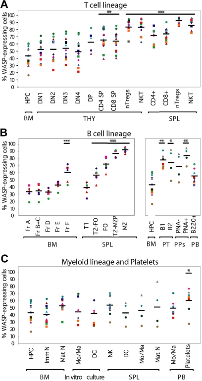
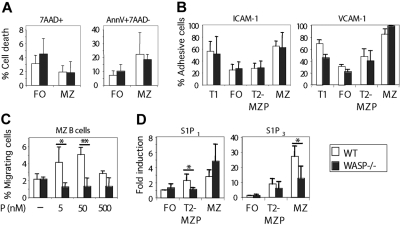
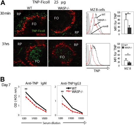
Development of hematopoietic cells depends on a dynamic actin cytoskeleton. Here we demonstrate that expression of the cytoskeletal regulator WASP, mutated in the Wiskott-Aldrich syndrome, provides selective advantage for the development of naturally occurring regulatory T cells, natural killer T cells, CD4(+) and CD8(+) T lymphocytes, marginal zone (MZ) B cells, MZ macrophages, and platelets. To define the relative contribution of MZ B cells and MZ macrophages for MZ development, we generated wild-type and WASP-deficient bone marrow chimeric mice, with full restoration of the MZ. However, even in the presence of MZ macrophages, only 10% of MZ B cells were of WASP-deficient origin. We show that WASP-deficient MZ B cells hyperproliferate in vivo and fail to respond to sphingosine-1-phosphate, a crucial chemoattractant for MZ B-cell positioning. Abnormalities of the MZ compartment in WASP(-/-) mice lead to aberrant uptake of Staphylococcus aureus and to a reduced immune response to TNP-Ficoll. Moreover, WASP-deficient mice have increased levels of "natural" IgM antibodies. Our findings reveal that WASP regulates both development and function of hematopoietic cells. We demonstrate that WASP deficiency leads to an aberrant MZ that may affect responses to blood-borne pathogens and peripheral B-cell tolerance.


Figures

Figure 1
Figure 1
Selective advantage of WASP-expressing cells in WASP+/− mice. HPCs and hematopoietic lineage cells were defined by surface and intracellular markers, labeled with anti-WASP antibodies, and analyzed by flow cytometry. Percentage of WASP-expressing cells is indicated in (A) the T-cell lineage, (B) the B-cell lineage, and (C) the myeloid lineage. Each color represents data from one mouse. Black bar represents the mean of the group. B indicates B cell; B1/B2, peritoneal B cells; BM, bone marrow; DC, dendritic cell; DN, CD4CD8 double-negative; DP, CD4CD8 double-positive; FO, follicular; Fr, fraction, HPC, hematopoietic progenitor cells; Mo/Ma, monocyte/macrophage; MZ, marginal zone; N, neutrophil; NK, natural killer cell; NKT, NK T cell; nTregs, naturally occurring T cells; PB, peripheral blood; PNA, peanut agglutinin; PPs, Peyer patches; PT, peritoneum; SP, CD4 or CD8 single-positive; SPL, spleen; T1/T2, transitional 1/2 B cell; T2-FO, transitional 2 follicular precursor B cell; T2-MZP, transitional 2 marginal zone precursor B cell; THY, thymus. In vitro cultured Mo/Ma and DCs were obtained by culturing BM cells in vitro for 7 days with M-CSF and GM-CSF, respectively. P values use a t test to compare percentage of WASP-expressing cells in the T-cell, B-cell (right panel), and myeloid lineages to that of HPCs and for the B-cell lineage (left panel) to that of Fr A (pro-B) cells: *P < .05, **P < .01, ***P < .005.
Figure 2
Figure 2
MZ B cells and MZMs are reduced, and S aureus uptake aberrant in WASP-deficient mice. Immunohistochemistry of spleen sections from WT and WASP−/− mice. (A) Schematic representation of a B-cell follicle and the MZ in the spleen. Black arrows indicate the MZ. (B) Reduced number of MZ B cells (CD1d+ B220+) and normal localization of metallophilic macrophages (MOMA-1+ Sialoadhesin+) in WASP−/− spleens relative to WT spleens. Black arrowhead represents the MZ. (C) Normal localization of red pulp macrophages (F4/80+), CD11b+ macrophages, and DCs (CD11c+). (D) Reduced number of MZMs (MARCO+ SIGN-R1+) in WASP−/− spleens compared with WT spleens. Black arrow represents MZMs in the MZ. (E) Localization of S aureus 30 minutes after intravenous injection. Note that S aureus almost exclusively colocalize with MARCO+ MZ macrophages in WT mice, whereas WASP−/− mice show aberrant localization of S aureus. White arrowhead represents the MZ. FO indicates B-cell follicle; MZ, marginal zone; RP, red pulp. Original magnifications: panels B to D, ×10; D, ×20.
Figure 3
Figure 3
WASP-deficient MZ B cells are reduced in number independent of MZMs. (A) Schematic representation of generation of BM chimeric mice. WT (expressing CD45.1) and WASP−/− (CD45.2) BM cells at a 1:3 ratio were injected into lethally irradiated WASP−/− recipient mice. Mice were analyzed 9 to 13 weeks after transplantation. Control mice received WT or WASP−/− BM alone. (B) Immunohistochemistry of spleen sections from mice receiving WT BM (left panel), WASP−/− BM (middle), and mixed WT:WASP−/− BM (right). Note that the MZ architecture in WT:WASP−/− BM chimeric mice is fully restored. (C) Splenocytes were labeled with anti-CD21, CD23, and IgM and analyzed by flow cytometry. (Top panel) The percentage of FO and MZ B cells in reconstituted mice is shown for the various conditions as illustrated in panel B. Note that WT:WASP−/− BM chimeric mice have fully restored MZ B-cell population (right). (Bottom panel) The percentage of WASP−/− CD45.2-expressing FO and MZ B cells is shown. Note the low proportion of WASP−/− MZ B cells in the WT:WASP−/− BM chimeric mice (right). The data are representative of 2 independent experiments, in which n = 7 mice were analyzed (original magnification ×10).
Figure 4
Figure 4
Peripheral B-cell development and homeostasis in WASP−/− mice. (A) Left panel; flow cytometric analysis of WT:WASP−/− BM chimeric mice to define the percentage of WASP+ (CD45.2) cells in the T1, FO, T2-MZP, and MZ B-cell populations, using CD21, CD23, and IgM as markers. The middle panel shows the results of one representative experiment, indicating (from top to bottom) the proportion of WASP+ (CD45.2) cells in T2-MZP, FO, MZ B cells, and T1 cells, respectively. The right panel shows the percentage of WASP-expressing (CD45.1+) cells in the indicated B-cell populations. The hatched line indicates the 25% of WASP+ BM cells that were used for transplantation. The right panel represents data from one of 2 similar experiments with n = 7 mice. (B) Proportion of T1, T2-FO, FO, T2-MZP, and MZ B cells in WT and WASP−/− mice (n = 10 mice per group). (C) Absolute numbers of T1, T2-FO, FO, T2-MZP, and MZ B cells in WT and WASP−/− mice (n = 10 mice per group). (D) In vivo BrdU labeling. (Top panel) Mice were fed BrdU in the drinking water for 6 days (pulse) and followed for another 6 days (chase). Proportion of BrdU positive cells was assessed at day 3, 6, 9, and 14. (Bottom panels) The proportion of BrdU+ cells of T1, T2-FO, FO, T2-MZP, and MZ B cells as defined by CD21, CD23, CD24, and IgM expression is shown (n = 4 mice per group). *P < .05, **P < .01, ***P < .005.
Figure 5
Figure 5
WASP-deficient MZ B cells show impaired in vitro migration to S1P. (A) Flow cytometric analysis of cell death in freshly isolated FO and MZ B cells, identified based on CD21 and CD23 expression. Necrotic cells were defined as 7-AAD+ (left panel) and apoptotic cells as 7-AAD annexin V+ (right). N = 6 mice per group. (B) Adhesive response of T1, FO, T2-MZP, and MZ B cells to ICAM-1 (left panel) and VCAM-1 (right). Splenocytes were allowed to adhere to tissue culture plates coated with indicated ligands for 2 hours. Adherent cells were released by ethylenediaminetetraacetic acid, stained for CD21, CD23, and IgM to define B-cell subsets, and enumerated by flow cytometry. The percentage of adhesive cells as a fraction of total input cells is shown as mean values (± SD). This experiment is representative of 2 similar ones with n = 3 mice per group. (C) Migratory response of MZ B cells to S1P was determined by an in vitro chemotaxis assay. The percentage of cells that migrated after 3 hours in the chemotaxis assay was determined and represents the mean value (± SD) of n = 3 mice per group. This experiment represents 2 similar experiments. (D) Real-time PCR analysis of S1P1, and S1P3 in FO, T2-MZP, and MZ B cells. B cells from 3 mice per group were pooled and cell-sorted based on CD21, CD23, and IgM expression. Samples were run in triplicate, and the target mRNA was normalized to HPRT mRNA. The mRNA content of test sample in WT FO B cells was defined as 1 arbitrary unit. Mean value (± SD) of triplicates is shown, and the data are representative of 3 similar experiments. *P < .05, **P < .01.
Figure 6
Figure 6
WASP-deficient mice show reduced specific immune response to TNP-Ficoll. WT and WASP−/− mice when 12 to 14 weeks old were injected intravenously with 2.5 μg of TNP-Ficoll. (A) Uptake of TNP-Ficoll in spleens 30 minutes and 3 hours after injection. (Left panel) Spleen sections were labeled to detect TNP-Ficoll and MOMA+ metallophilic macrophages to define the MZ. Note the reduced TNP staining in the MZ at 30 minutes (top panels) and in the follicles at 3 hours (bottom panels) in WASP−/− mice compared with WT mice (original magnification ×20). (Right panels) MZ B cells were labeled with anti-TNP and analyzed by flow cytometry (n = 5). (B) Anti-TNP IgM and IgG3 antibody titers were determined at day 7 after immunization by enzyme-linked immunosorbent assay. Each group represents mean values (± SD) from n = 5 mice, which were corrected for background binding. FO indicates B-cell follicle; MZ, marginal zone; non-B, lymphocytes negative for CD21 and IgM; RP, red pulp. *P < .05, **P < .01.

Comment in

  • WASp stings mature lymphocytes.
    Severinson E. Severinson E. Blood. 2008 Nov 15;112(10):3921-2. doi: 10.1182/blood-2008-09-176685. Blood. 2008. PMID: 18988873 Free PMC article.

References

    1. Notarangelo LD, Miao CH, Ochs HD. Wiskott-Aldrich syndrome. Curr Opin Hematol. 2008;15:30–36. - PubMed
    1. Filipovich AH, Stone JV, Tomany SC, et al. Impact of donor type on outcome of bone marrow transplantation for Wiskott-Aldrich syndrome: collaborative study of the International Bone Marrow Transplant Registry and the National Marrow Donor Program. Blood. 2001;97:1598–1603. - PubMed
    1. Pai SY, DeMartiis D, Forino C, et al. Stem cell transplantation for the Wiskott-Aldrich syndrome: a single-center experience confirms efficacy of matched unrelated donor transplantation. Bone Marrow Transplant. 2006;38:671–679. - PubMed
    1. Ozsahin H, Cavazzana-Calvo M, Notarangelo LD, et al. Long-term outcome following hematopoietic stem-cell transplantation in Wiskott-Aldrich syndrome: collaborative study of the European Society for Immunodeficiencies and European Group for Blood and Marrow Transplantation. Blood. 2008;111:439–445. - PubMed
    1. Dewey RA, Avedillo Diez I, Ballmaier M, et al. Retroviral WASP gene transfer into human hematopoietic stem cells reconstitutes the actin cytoskeleton in myeloid progeny cells differentiated in vitro. Exp Hematol. 2006;34:1161–1169. - PubMed

Publication types

MeSH terms