. 2008;3(9):1402-13.
doi: 10.1038/nprot.2008.120.

Evaluating cell-surface expression and measuring activation of mammalian odorant receptors in heterologous cells

Affiliations

Evaluating cell-surface expression and measuring activation of mammalian odorant receptors in heterologous cells

Hanyi Zhuang et al. Nat Protoc. 2008.

Abstract

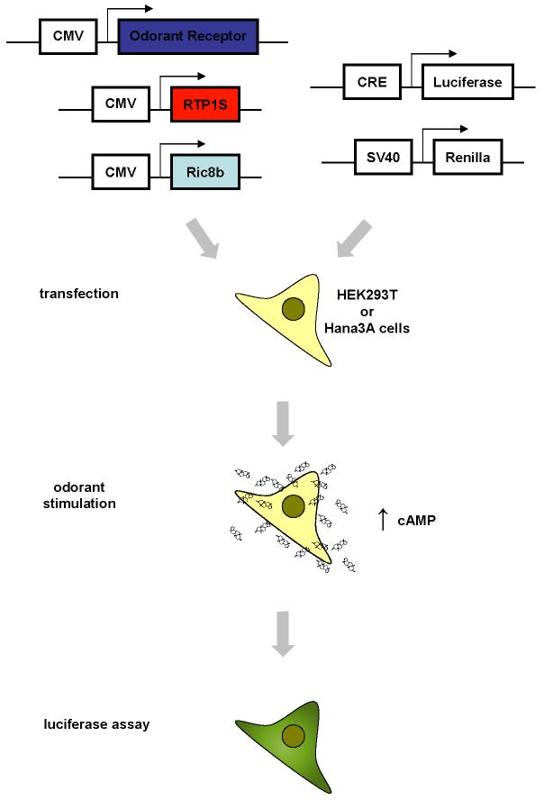
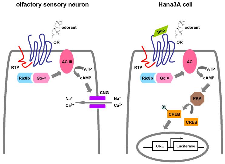
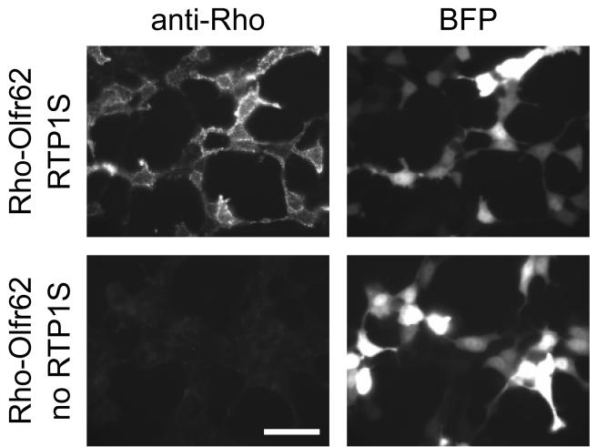
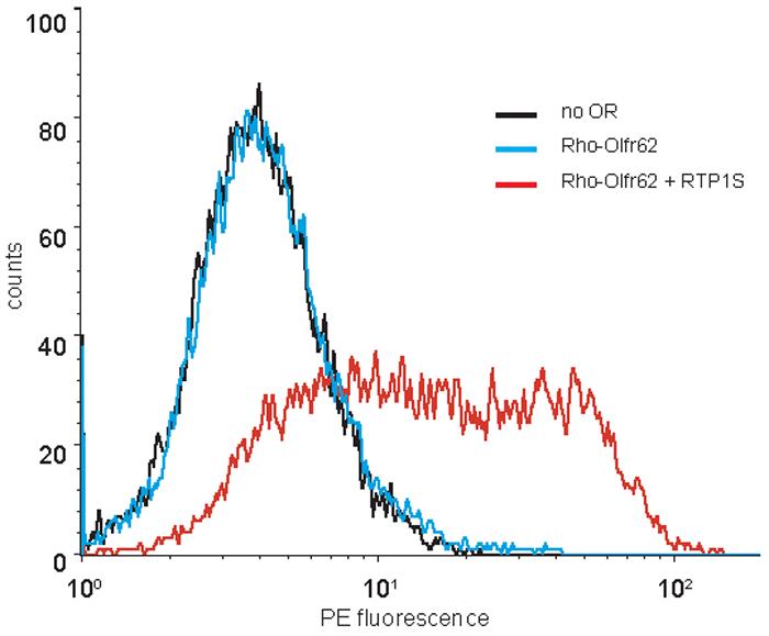
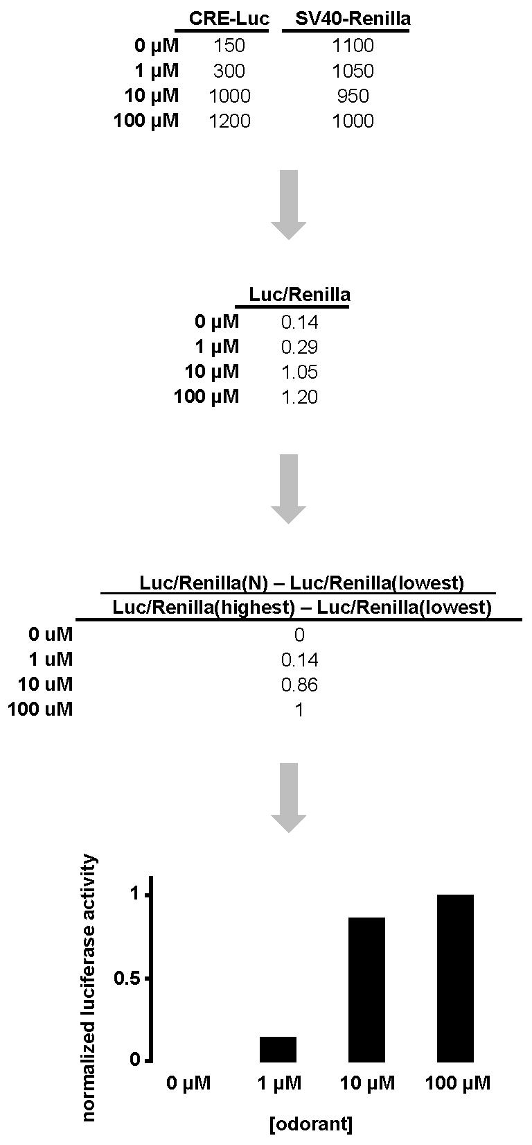
A fundamental question in olfaction is which odorant receptors (ORs) are activated by a given odorant. A major roadblock to investigating odorant-OR relationships in mammals has been the inability to express ORs in heterologous cells suitable for screening active ligands for ORs. The discovery of the receptor-transporting protein family has facilitated the effective cell-surface expression of ORs in heterologous cells. The establishment of a robust heterologous expression system for mammalian ORs facilitates the high-throughput 'deorphanization' of these receptors by matching them to their cognate ligands. This protocol details the method used for evaluating the cell-surface expression and measuring the functional activation of ORs of transiently expressed mammalian ORs in HEK293T cells. The stages of OR cell-surface expression include cell culture preparation, transfer of cells, transfection, immunocytochemistry or flow cytometry, odorant stimulation and luciferase assay. This protocol can be completed in a period of 3 d from the transfer of cells to cell-surface expression detection and/or measurement of functional activation.


Figures

Figure 1
Figure 1. Overview of the functional activation of odorant receptors in a heterologous expression system
A mammalian odorant receptor, RTP1S, and Ric8b, each contained in a mammalian expression vector such as pCI, are transfected into HEK293T or the HEK293T-derived Hana3A cells along with a luciferase reporter gene construct driven by a cyclic AMP responsive element (CRE) promoter and Renilla luciferase construct drive by an SV40 promoter. Typically one day after transfection, cells are stimulated with potential odorants. Activation of the odorant receptor leads to accumulation of cyclic AMP (cAMP) which turns on the expression of the luciferase reporter gene.
Figure 2
Figure 2. The endogenous and heterologous odorant receptor signal transduction pathways
A) The endogenous odorant receptor signal transduction pathway in an olfactory sensory neuron. B) The heterologous odorant receptor signal transduction pathway in Hana3A cells. OR, odorant receptor. RTP, receptor-transporting protein. PKA, protein kinase A. CREB, cAMP response element binding protein. CRE, cAMP response element.
Figure 3
Figure 3
An example of live-cell immunocytochemistry staining setup with antibody solution on two samples
Figure 4
Figure 4. An example of cell-surface odorant receptor expression
Rho-Olfr62 is cotransfected with (upper panels) and without (lower panels) RTP1S. Robust Olfr62 expression is seen as punctate signals when RTP1S is cotransfected and cells are stained with anti-Rhodopsin antibody (top left hand side panel). Lack of cell-surface expression is seen when no RTP1S is cotransfected (bottom left hand side panel). Blue fluorescent protein (BFP) is cotransfected as a control for transfection efficiency (right hand side panels). Scale bar = 50 μm.
Figure 5
Figure 5. An example of cell-surface flow cytometry
The intensity of the phycoerythrin (PE) signal among the 10,000 GFP-positive cells is measured and plotted. Rho-Olfr62 is cotransfected with and without RTP1S. Robust Olfr62 expression is seen as right-shifted plots when RTP1S is cotransfected and cells are stained with anti-Rhodopsin antibody (red line). Lack of cell-surface expression is seen when no RTP1S is cotransfected (compare blue line and negative control black line).
Figure 6
Figure 6. A hypothetical case of luciferase assay data interpretation
CRE-mediated firefly luciferase (CRE-Luc) measurements and SV40-mediated Renilla luciferase (SV40-Renilla) measurements are obtained for a hypothetical odorant receptor tested against 100 μM, 10 μM, 1 μM, and 0 μM (negative control) of a hypothetical odorant. The ratio of firefly luciferase and Renilla luciferase (Luc/Renilla) adjusts for difference in cell number and transfection efficiency among experiments. Normalization of the data is calculated by the formula [Luc/Renilla(N) - Luc/Renilla(lowest)] / [Luc/Renilla(highest) - Luc/Renilla(lowest)]. A column graph is drawn based on the normalized luciferase activity at each concentration.

References

    1. Barnea G, et al. Odorant receptors on axon termini in the brain. Science. 2004;304:1468. - PubMed
    1. Strotmann J, Levai O, Fleischer J, Schwarzenbacher K, Breer H. Olfactory receptor proteins in axonal processes of chemosensory neurons. J Neurosci. 2004;24:7754–61. - PMC - PubMed
    1. Niimura Y, Nei M. Extensive gains and losses of olfactory receptor genes in Mammalian evolution. PLoS ONE. 2007;2:e708. - PMC - PubMed
    1. Niimura Y, Nei M. Evolutionary changes of the number of olfactory receptor genes in the human and mouse lineages. Gene. 2005;346:23–8. - PubMed
    1. Godfrey PA, Malnic B, Buck LB. The mouse olfactory receptor gene family. Proc Natl Acad Sci U S A. 2004;101:2156–61. - PMC - PubMed

Publication types