. 2008 Dec;15(12):1921-9.
doi: 10.1038/cdd.2008.127. Epub 2008 Sep 5.

NRAGE, a p75NTR adaptor protein, is required for developmental apoptosis in vivo

Affiliations

NRAGE, a p75NTR adaptor protein, is required for developmental apoptosis in vivo

M J M Bertrand et al. Cell Death Differ. 2008 Dec.

Abstract

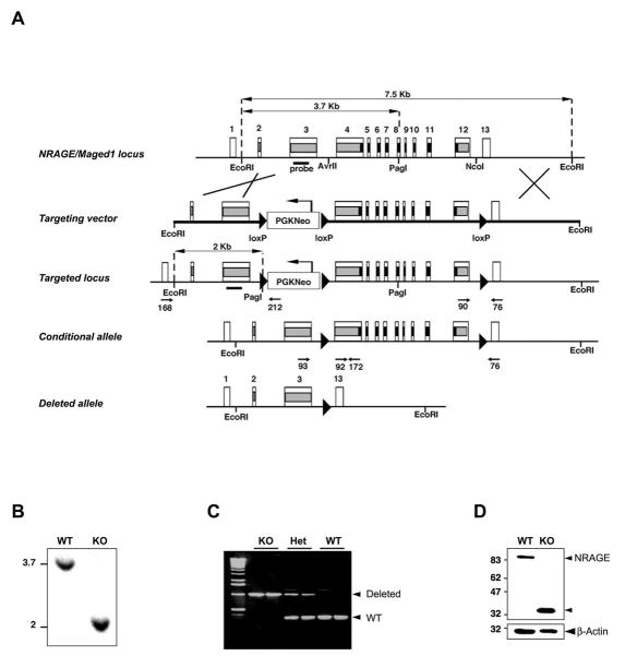
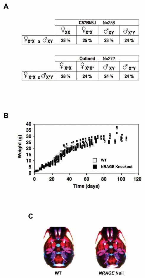
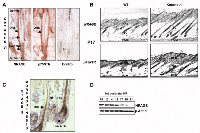
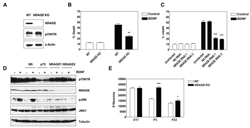
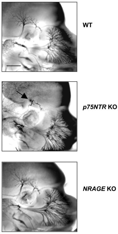
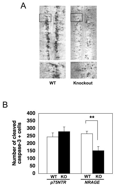
NRAGE (also known as Maged1, Dlxin) is a member of the MAGE gene family that may play a role in the neuronal apoptosis that is regulated by the p75 neurotrophin receptor (p75NTR). To test this hypothesis in vivo, we generated NRAGE knockout mice and found that NRAGE deletion caused a defect in developmental apoptosis of sympathetic neurons of the superior cervical ganglia, similar to that observed in p75NTR knockout mice. Primary sympathetic neurons derived from NRAGE knockout mice were resistant to apoptosis induced by brain-derived neurotrophic factor (BDNF), a pro-apoptotic p75NTR ligand, and NRAGE-deficient sympathetic neurons show attenuated BDNF-dependent JNK activation. Hair follicle catagen is an apoptosis-like process that is dependent on p75NTR signaling; we show that NRAGE and p75NTR show regulated co-expression in the hair follicle and that identical defects in hair follicle catagen are present in NRAGE and p75NTR knockout mice. Interestingly, NRAGE knockout mice have severe defects in motoneuron apoptosis that are not observed in p75NTR knockout animals, raising the possibility that NRAGE may facilitate apoptosis induced by receptors other than p75NTR. Together, these studies demonstrate that NRAGE plays an important role in apoptotic-signaling in vivo.


Figures

Figure 1
Figure 1. Targeted mutation of NRAGE/Maged1
(A) Genomic organization of the NRAGE/Maged1 locus and schematic representation of the inactivation procedure. The conditional allele (Maged1tm1Urfm) and the deleted allele (Maged1tm1.1Urfm), generated after Cre-mediated recombination, are shown. Filled boxes represent the coding region of NRAGE, the segment encoding the p75NTR binding domain is in black. The loxP sites are represented by solid triangles. (B) Southern blot analysis of DNA isolated from wild-type ES cells and from ES cells that have undergone gene replacement (EcoRI/PagI double digestion). (C) PCR analysis of DNA isolated from tail biopsies of NRAGE knockout, heterozygous and wild-type mice. PCR amplification with primers 93 and 76 gives rise to a 940-bp product specific of the deleted allele (Maged1tm1.1Urfm) while amplification with primers 92 and 172 produces a 270-bp fragment specific of the wild-type allele. (D) Immunoblot blot analysis of protein lysates from NRAGE knockout and wild-type E13.5 brains.
Figure 2
Figure 2. NRAGE deficiency has no impact on viability
(A) The fraction of each genotype generated by crossing heterozygous females (X*X) with either wild-type (XY) or hemizygous (X*Y) males do not differ from Mendelian predictions on either a pure C57Bl/6 background or in outbred CD1 strains. (B) NRAGE knockout and wild-type littermates display similar weight gain. (C) Alcian blue blue/alizarin red staining of P0 mice did not reveal abnormalities in skeletal development.
Figure 3
Figure 3. Hair follicle catagen is retarded in NRAGE knockout mice
(A) Immunocytochemistry performed on serial back skin sections of a P18 C57BL/6 mouse showing expression of NRAGE and p75NTR protein in the outer root sheath of a hair follicle on catagen VI. (B) Retardation of hair follicle regression in P17 NRAGE knockout mice resembles that observed in P17 p75NTR knockout mice. Note the thickening of the dermis in each of the null strains. (C) In situ hybridization using an NRAGE probe labeled with digoxygenin showing NRAGE RNA expression in the inner root sheath, the outer root sheath and hair bulb within a hair follicle of a P4 C57BL/6 mouse. (D) NRAGE protein expression at different stages of the first hair follicle cycle. Scale bar in B is 50 μm. IRS, inner root sheath; ORS, outer root sheath; DER, dermis; SC, subcutis; PCM, panniculus carnosus muscle.
Figure 4
Figure 4. NRAGE knockout SCG sympathetic neurons are deficient in p75NTR-induced apoptosis
(A) NRAGE and p75NTR expression in lysates from P4 SCGs of NRAGE knockout and wild-type control pups was determined by immunoblot. (B) Sympathetic neurons derived from NRAGE null animals or from wild-type littermates were exposed to BDNF and apoptotic nuclei were quantified 48 hours later. Approximately 50–70 neurons were counted for each condition. The experiment was performed in triplicate. **: P=0.0022. (C) Rat sympathetic neurons were infected with lentivirus expressing miRNA that targets NRAGE or were infected with a negative control lentivirus expressing miRNA that does not target a mammalian protein (NS RNAi), then cultured for 5 days. Neurons were then exposed to BDNF for 72 hours and apoptotic nuclei were quantified. ***: P<0.0001. (D) Rat sympathetic neurons were infected as in (C) but were also infected with a lentivirus expressing miRNA that targets p75NTR. 6 days later, neurons were exposed to BDNF for 1 hour, lysed and analyzed by immunoblot, as indicated. (E) Number of neurons within the superior cervical ganglia were quantified in NRAGE null and wild-type littermates at E17 (n=3), at P3 (n=5) and at P23 (NRAGE knockout: n=8, wild-type controls: n=10). ***: P<0.0001, *: P=0.05. For B, C and E, significance values were calculated using unpaired student t tests. Error bars represent SEM.
Figure 5
Figure 5. Abnormal outgrowth of the ophthalmic branch of the trigeminal ganglion in p75NTR knockout mice but not in NRAGE knockout mice
Whole-mount immunostaining of wild-type, p75NTR knockout and NRAGE knockout embryos with TuJ1, a monoclonal antibody raised against the neuron specific marker β3 tubulin. Arrow indicates growth defect in the ophthalmic branch of the trigeminal ganglion in p75NTR knockout embryos. Scale bar: 0.35 mm.
Figure 6
Figure 6. NRAGE, but not p75NTR, is required for motoneuron apoptosis in vivo
(A) Whole-mount cleaved caspase-3 immunostaining of the lumbar region of E13.5 spinal cords from wild-type and NRAGE knockout embryos. Bottom panels represent magnifications of the inset regions. (B) Quantification of lumbar spinal motoneuron apoptosis. NRAGE knockout: n=4, wild-type littermates: n=5; p75NTR knockout: n=8, wild-type littermates: n=12. **: P=0.0081. P value was calculated using unpaired, two-tailed student t test. Error bars represent s.e.m

References

    1. Roux PP, Barker PA. Neurotrophin signaling through the p75 neurotrophin receptor. Prog Neurobiol. 2002;67:203–33. - PubMed
    1. Nykjaer A, Willnow TE, Petersen CM. p75NTR--live or let die. Curr Opin Neurobiol. 2005;15:49–57. - PubMed
    1. Teng KK, Hempstead BL. Neurotrophins and their receptors: signaling trios in complex biological systems. Cell Mol Life Sci. 2004;61:35–48. - PMC - PubMed
    1. Beattie MS, Harrington AW, Lee R, Kim JY, Boyce SL, Longo FM, et al. ProNGF induces p75-mediated death of oligodendrocytes following spinal cord injury. Neuron. 2002;36:375–86. - PMC - PubMed
    1. Bhakar AL, Howell JL, Paul CE, Salehi AH, Becker EB, Said F, et al. Apoptosis induced by p75NTR overexpression requires Jun kinase-dependent phosphorylation of Bad. J Neurosci. 2003;23:11373–81. - PMC - PubMed

Publication types

Substances