. 2008 Sep 5;4(9):e1000174.
doi: 10.1371/journal.pgen.1000174.

Identification of neural crest and glial enhancers at the mouse Sox10 locus through transgenesis in zebrafish

Affiliations

Identification of neural crest and glial enhancers at the mouse Sox10 locus through transgenesis in zebrafish

Anthony Antonellis et al. PLoS Genet. .

Abstract

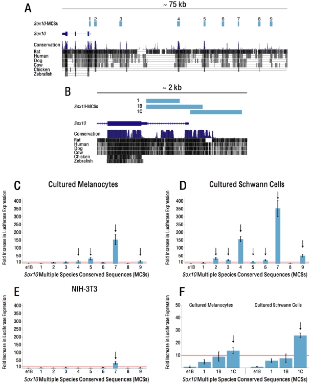
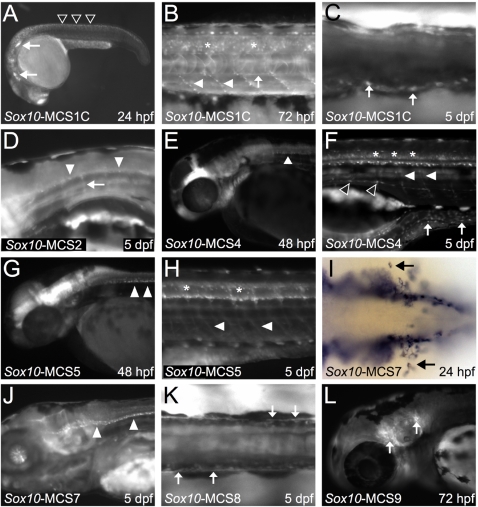
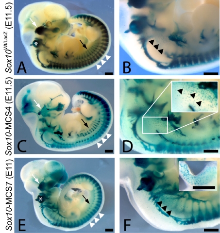
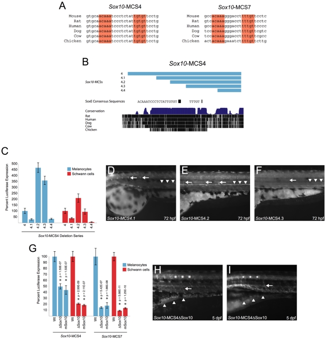
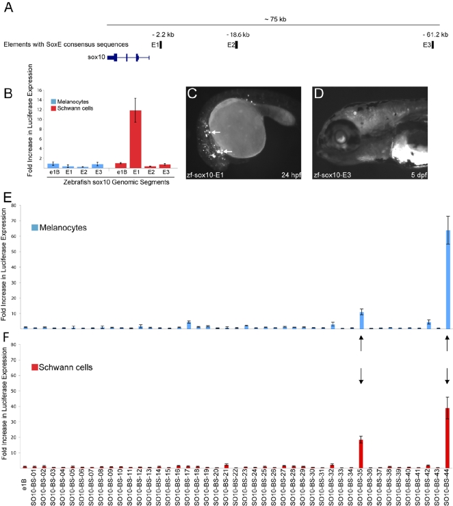
Sox10 is a dynamically regulated transcription factor gene that is essential for the development of neural crest-derived and oligodendroglial populations. Developmental genes often require multiple regulatory sequences that integrate discrete and overlapping functions to coordinate their expression. To identify Sox10 cis-regulatory elements, we integrated multiple model systems, including cell-based screens and transposon-mediated transgensis in zebrafish, to scrutinize mammalian conserved, noncoding genomic segments at the mouse Sox10 locus. We demonstrate that eight of 11 Sox10 genomic elements direct reporter gene expression in transgenic zebrafish similar to patterns observed in transgenic mice, despite an absence of observable sequence conservation between mice and zebrafish. Multiple segments direct expression in overlapping populations of neural crest derivatives and glial cells, ranging from pan-Sox10 and pan-neural crest regulatory control to the modulation of expression in subpopulations of Sox10-expressing cells, including developing melanocytes and Schwann cells. Several sequences demonstrate overlapping spatial control, yet direct expression in incompletely overlapping developmental intervals. We were able to partially explain neural crest expression patterns by the presence of head to head SoxE family binding sites within two of the elements. Moreover, we were able to use this transcription factor binding site signature to identify the corresponding zebrafish enhancers in the absence of overall sequence homology. We demonstrate the utility of zebrafish transgenesis as a high-fidelity surrogate in the dissection of mammalian gene regulation, especially those with dynamically controlled developmental expression.


Conflict of interest statement

The authors have declared that no competing interests exist.

Figures

Figure 1
Figure 1. Sox10-MCSs display enhancer activity in Neural Crest–derived cell lines.
A) Sox10-MCSs (pale blue bars) identified within and upstream of the Sox10 locus, depicted by a 75 kb interval from the UCSC Genome Browser (genome.ucsc.edu). B) Enlargement of a 2 kb interval encompassing the Sox10 transcriptional start site. Three elements, comprising sequence from within intron one alone (Sox10-MCS1), intron one and sequence 5′ of the TSS (Sox10-MCS1B), and sequence only 5′ of the TSS (Sox10-MCS1C) were subcloned and tested individually for enhancer activity. C–F) Sox10-MCSs cloned upstream of a minimal promoter driving luciferase reporter expression were assayed for enhancer activity in melan-a cells (C and F), S16 cells (D and F), and NIH-3T3 cells (E). The red line demarcates the 10-fold cut-off for “enhancer activity” (see Results); arrows indicate sequences exceeding that activity threshold. Error bars, SD (standard deviation).
Figure 2
Figure 2. Mouse Sox10-MCSs direct EGFP reporter expression in zebrafish embryos consistent with endogenous sox10.
A–C) Sox10-MCS1C directs reporter gene expression to the cranial ganglia (A; white arrows) and within premigratory neural crest (A; open arrowheads) at 24 hpf (A). By 72 hpf (B), signal is detected in scattered oligodendrocytes (asterisks) along the spinal column and descending Schwann cells (white arrowheads) surrounding peripheral motor neurons. Weak reporter expression is also detected along the sympathetic chain (B; white arrow). At 5 dpf (C), signal is clearly detected in the ENS (C; white arrows). D) Sox10-MCS2 directs weak EGFP expression in glial cells of the CNS (white arrowheads) and PNS (white arrow). E and F) Sox10-MCS4 directs reporter gene expression in presumptive oligodendroglial cells at 48 hpf (E; white arrowhead). By 5 dpf (F), robust signal is detected in several neural crest lineages, including Schwann cells (F; white arrowheads), sympathetic chain ganglia (open arrowheads), and the enteric nervous system (white arrows). As with endogenous sox10, EGFP expression is maintained in mature oligodendrocytes (asterisks). G and H) Sox10-MSC5 also directs signal in early oligodendroglial cells (G; white arrowheads) and sustains signal in mature oligodendrocytes (asterisks). Glial cells of the PNS (H; white arrowheads) are clearly detected at 5 dpf (H). I) In situ hybridization using GFP riboprobe detects early migrating melanoblasts (black arrows) in Sox10-MCS7 transgenic embryos at 24 hpf. J) By 5 dpf, Sox10-MCS7 directs signal to oligodendrocytes (white arrowheads) along the ventral column. K) Sox10-MCS8 directs weak reporter to the ENS (white arrows) at 5 dpf. L) at 72 hpf, sustained reporter gene expression appears in the cranial ganglia (white arrows) of Sox10-MCS9 transgenic zebrafish embryos.
Figure 3
Figure 3. Summary of regulatory activities displayed by Sox10-MCSs in cultured cells and in developing zebrafish embryos.
A schematic of the assayed Sox10-MCS segments is depicted at the top. Distance in kb from the Sox10 TSS is shown in the brackets adjacent to each construct. Expression in cultured cells and zebrafish cell populations is noted below each construct. For the cultured cells, black-filled diamonds denote at least a 10-fold enhancement in our in vitro analysis, while white-filled diamonds fail to reach that threshold. For the neural crest–derived populations and oligodendrocytes evaluated in transgenic zebrafish embryos, black-filled diamonds refer to strong reporter expression, grey-filled diamonds to weaker expression, and white-filled diamonds to an undetected level of reporter expression.
Figure 4
Figure 4. Sox10-MCS4 and Sox10-MCS7 direct reporter expression in developing transgenic mice consistent with their activity in zebrafish.
LacZ reporter expression was detected at the resolution of whole-mount for Sox10 Wt/tm1Weg embryos at E11.5 (A and B), Sox10-MCS4 embryos at E11.5 (C and D), and Sox10-MCS7 embryos at E11 (E and F). The broad expression of Sox10 in neural crest–derived populations is apparent in Sox10 Wt/tm1Weg embryos (A and B), including the cranial ganglia (white arrow), otic vesicle (asterisk), sympathetic ganglia (black arrow), dorsal root ganglia (white arrowheads), and melanoblasts (black arrowheads). Both Sox10-MCS4 (C and D) and Sox10-MCS7 (E and F) also direct similarly broad reporter expression in these neural crest tissues. D and F insets represent melanoblast and enteric ganglia expression, respectively. Scale bar = 500 µm.
Figure 5
Figure 5. Analyses of a dimeric SoxE consensus sequence within Sox10-MCS4 and Sox10-MCS7.
A) Consensus SoxE family binding sites are oriented in a head-to-head fashion within Sox10-MCS4 and Sox10-MCS7. B) A deletion series across Sox10-MCS4 (pale blue bars) is depicted in the UCSC Genome Browser. The position and sequence of a monomeric and a dimeric SoxE consensus sequence is shown below the deletion series. C) In vitro enhancer activity for each fragment of the deletion series was tested individually in melan-a cells (blue bars) and S16 cells (red bars). The results for Sox10-MCS4 are included for cross-comparison of modular enhancer activity. Error bars, SD. D–F) in vivo enhancer activity was compared at 72 hpf for Sox10-MCS4.1 through Sox10-MCS4.3 in transgenic zebrafish embryos. Sox10-MCS4.4 failed to direct reporter expression in G0 embryos and was not raised for germline transmission. Sox10-MCS4.1 (D) directed reporter expression to Schwann cells (white arrows) and weak reporter expression to sympathetic ganglia (white arrowheads). Sox10-MCS4.2 (E) directed reporter expression in an opposite fashion, as signal appeared weaker in Schwann cells and more robust in sympathetic ganglia. Sox10-MCS4.3 (F) directed an extremely low level of reporter expression to these two neural crest–derived populations, and the arrow and arrowheads show the relative position of where the Schwann cells and sympathetic ganglia is normally positioned. G) Site-directed mutagenesis was used to delete and mutate the dimeric SoxE consensus sequence within Sox10-MCS4 and Sox10-MCS7. These mutagenized constructs were tested for in vitro enhancer activity in melanocytes (blue bars) and Schwann cells (red bars), and compared against their wild-type sequences. P-values are given above each tested construct and error bars indicate the standard deviation. H and I) Sox10-MCS4 with a deleted head-to-head SoxE family binding site was selected for transmission through the germline. Analysis of two different founder lines (H and I, respectively) revealed a decrease in signal in oligodendrocytes (asterisks) and scattered reporter expression in a subset of the ENS (white arrowheads). Schwann cells (white arrow), however, appear to be unaffected.
Figure 6
Figure 6. Motif-based search for functional zebrafish orthologs and SoxE positive enhancers.
A) A computational search for dimeric SoxE consensus sequences in a 100 kb window upstream of the zebrafish sox10 gene resulted in three hits (pale blue bars). The distance from the sox10 TSS is denoted above each enhancer (E1–E3). B) A 500 bp region surrounding each dimeric SoxE consensus sequence was tested for in vitro enhancer activity in a melanocyte cell line (blue bars) and a Schwann cell line (red bars). Each fragment is compared against a control (pE1b with no insert) and error bars indicate the standard deviation. C) zf-sox10-E1 directs reporter expression in scattered cells throughout the 24 hpf, G0 embryo, consistent with migrating neural crest cells (white arrows). D) zf-sox10-E3 directs reporter expression in cranial oligodendrocytes (asterisks) at 5 dpf in G0 embryos. E and F) Data mining of the mouse genome for highly conserved dimeric SoxE consensus sequences in the intron of genes expressed in melanocytes identified 44 genomic segments. In vivo enhancer activity was tested in cultured melanocytes (E) and Schwann cells (F). Only two segments drove luciferase expression above a 10-fold threshold in both cell lines (black arrows). Error bars, SD.

Similar articles

Cited by

References

    1. Kalcheim C, Le Douarin NM. Requirement of a neural tube signal for the differentiation of neural crest cells into dorsal root ganglia. Dev Biol. 1986;116:451–466. - PubMed
    1. Bondurand N, Kobetz A, Pingault V, Lemort N, Encha-Razavi F, et al. Expression of the SOX10 gene during human development. FEBS Lett. 1998;432:168–172. - PubMed
    1. Pusch C, Hustert E, Pfeifer D, Sudbeck P, Kist R, et al. The SOX10/Sox10 gene from human and mouse: sequence, expression, and transactivation by the encoded HMG domain transcription factor. Hum Genet. 1998;103:115–123. - PubMed
    1. Herbarth B, Pingault V, Bondurand N, Kuhlbrodt K, Hermans-Borgmeyer I, et al. Mutation of the Sry-related Sox10 gene in Dominant megacolon, a mouse model for human Hirschsprung disease. Proc Natl Acad Sci U S A. 1998;95:5161–5165. - PMC - PubMed
    1. Kapur RP. Early death of neural crest cells is responsible for total enteric aganglionosis in Sox10(Dom)/Sox10(Dom) mouse embryos. Pediatr Dev Pathol. 1999;2:559–569. - PubMed

Publication types

MeSH terms