. 2008 Sep 5;4(9):e1000177.
doi: 10.1371/journal.pgen.1000177.

A Sox10 expression screen identifies an amino acid essential for Erbb3 function

Affiliations

A Sox10 expression screen identifies an amino acid essential for Erbb3 function

Kristina Buac et al. PLoS Genet. .

Abstract

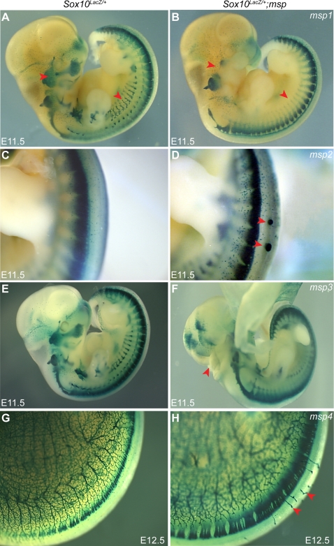
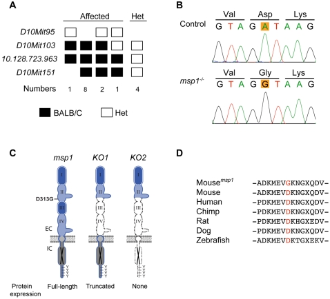
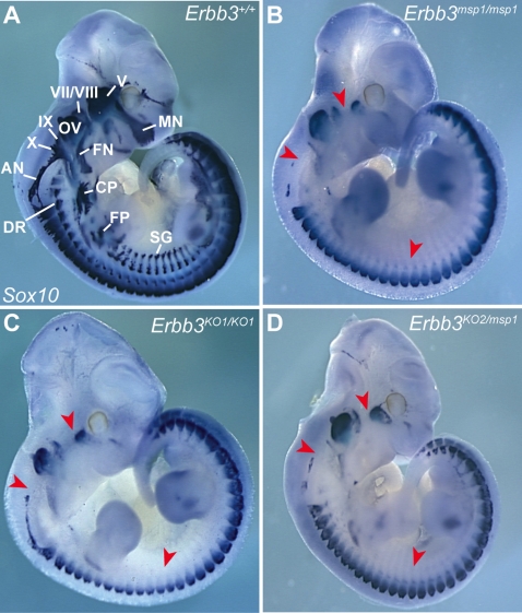
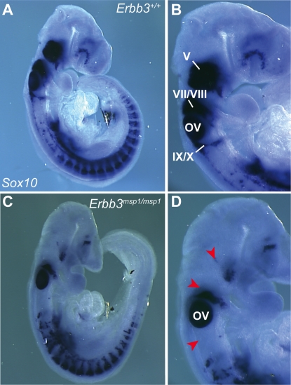
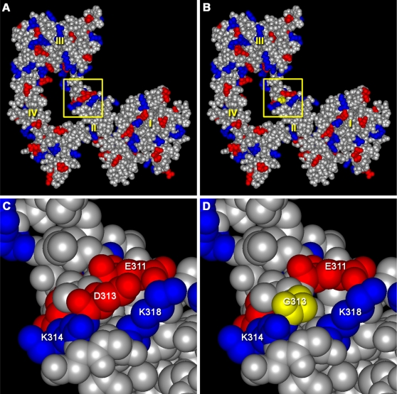
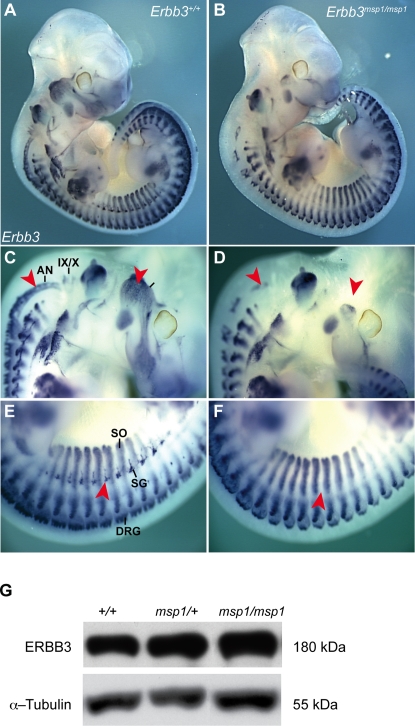
The neural crest (NC) is a population of embryonic stem cells that gives rise to numerous cell types, including the glia and neurons of the peripheral and enteric nervous systems and the melanocytes of the skin and hair. Mutations in genes and genetic pathways regulating NC development lead to a wide spectrum of human developmental disorders collectively called neurocristopathies. To identify molecular pathways regulating NC development and to understand how alterations in these processes lead to disease, we established an N-ethyl-N-nitrosourea (ENU) mutagenesis screen utilizing a mouse model sensitized for NC defects, Sox10(LacZ/+). Out of 71 pedigrees analyzed, we identified and mapped four heritable loci, called modifier of Sox10 expression pattern 1-4 (msp1-4), which show altered NC patterning. In homozygous msp1 embryos, Sox10(LacZ) expression is absent in cranial ganglia, cranial nerves, and the sympathetic chain; however, the development of other Sox10-expressing cells appears unaffected by the mutation. Linkage analysis, sequencing, and complementation testing confirmed that msp1 is a new allele of the receptor tyrosine kinase Erbb3, Erbb3(msp1), that carries a single amino acid substitution in the extracellular region of the protein. The ENU-induced mutation does not alter protein expression, however, it is sufficient to impair ERBB3 signaling such that the embryonic defects observed in msp1 resemble those of Erbb3 null alleles. Biochemical analysis of the mutant protein showed that ERBB3 is expressed on the cell surface, but its ligand-induced phosphorylation is dramatically reduced by the msp1 mutation. These findings highlight the importance of the mutated residue for ERBB3 receptor function and activation. This study underscores the utility of using an ENU mutagenesis to identify genetic pathways regulating NC development and to dissect the roles of discrete protein domains, both of which contribute to a better understanding of gene function in a cellular and developmental setting.


Conflict of interest statement

The authors have declared that no competing interests exist.

Figures

Figure 1
Figure 1. ENU Mutagenesis Screen Identifies Four Embryonic Phenotypes Affecting Sox10LacZ-Expression Pattern.
An age-matched wild-type control embryo (A, C, E, G) is shown next to a representative homozygote embryo for msp1 (B), msp2 (D), msp3 (F), and msp4 (H). The Sox10LacZ/+; msp1−/− embryos show reduction in Sox10LacZ- expressing cells in the cranial ganglia, cranial nerves and the sympathetic ganglia (B) (red arrowheads). Note the ectopic Sox10LacZ-expressing masses along the dorsal surface of the neural tube in Sox10LacZ/+; msp2−/− embryos (D) (red arrowheads). Neural tube fails to close in Sox10LacZ/+; msp3−/− embryos (F) (red arrowheads), thus disrupting normal Sox10LacZ expression in hindbrain. The Sox10LacZ/+; msp4−/− embryos show aberrant Sox10LacZ-expressing projections (H) (red arrowheads).
Figure 2
Figure 2. Linkage Analysis and Molecular Characterization of the msp1 Mutation.
(A) Fine mapping of the msp1 locus on mouse Chr 10. The mutation is confined within a region distal to D10Mit103. The number of affected mice analyzed is shown below genotyping data (homozygote black square; heterozygote white square). (B) Sequencing results of Erbb3 from control and msp1 embryos. The msp1 embryos carry an A-to-G transition in exon 8 of the Erbb3 gene that results in change of the aspartic acid (D) to glycine (G) at position 313. (C) Position of the msp1 mutation (D313G) in the ERBB3 protein. The ERBB3 protein consists of an extracellular region (EC), a single transmembrane-spanning region, and an intracellular (IC) portion that contains a conserved tyrosine-kinase domain flanked by a carboxy-terminal tail with tyrosine phosphorylation sites. The extracellular region is further subdivided into four domains: domain I, II, III, and IV. Domains I and III are homologous; domains II and IV are homologous. ERBB3 is a kinase-deficient receptor (black X). The D313G is located in the domain II (also known as cysteine-rich region I (CR1)) of ERBB3. In Erbb3KO1 allele a large portion of the extracellular domain is deleted, thus forming a truncated ERBB3 protein. In Erbb3KO2 allele no protein is made due to the insertion of premature termination codons in all three reading frames. (D) Multiple sequence alignment of Erbb3 from various species. The substituted aspartic acid (D313) is highly conserved among different species.
Figure 3
Figure 3. Complementation Test between msp1 and Erbb3 Null Allele in Mice.
Heterozygous msp1/+ mice were mated with Erbb3KO1/+ and Erbb3K02/+ mice to generate compound heterozygote embryos. Sox10 expression was analyzed in control (A), msp1 (B), null (C), and compound heterozygous embryos (D) at E11.5 by in situ hybridization. The mutant genotypes tested showed reduced expression of the Sox10 transcript in the cranial ganglia, cranial nerves and sympathetic ganglia (red arrowheads). Lack of complementation of Sox10 expression pattern in the compound heterozygous embryos confirmed that msp1 is allelic to Erbb3. The Sox10-expressing structures in the Erbb3+/+ embryo are: maxillary nerve (MN), facial nerve (FN), cervical plexus (CP), forelimb plexus (FP), dorsal ramus (DR), sympathetic ganglia (SG), trigeminal ganglia (V), geniculate ganglia (VII), acoustic ganglia (VIII), superior ganglia (IX), jugular ganglia (X), accessory nerve (AN) and otic vesicle (OV).
Figure 4
Figure 4. Expression Analysis of Endogenous Sox10 Transcript at E9.0.
In Erbb3+/+ (A, B), Sox10-expressing cells are found in the cranial ganglia and DRGs. In Erbb3msp1/msp1 (C, D), Sox10-expressing cells are detected in DRG but reduced in the cranial ganglia (red arrowheads). Labeled are trigeminal ganglia (V), geniculate and acoustic ganglia (VII/VIII), superior and jugular ganglia (IX/X) and the otic vesicle (OV).
Figure 5
Figure 5. Modeling the msp1 Mutation.
Structural models of mouse wild type (A) and msp1-mutant (B) ERBB3 extracellular domains in the “closed” conformation. Negatively charged residues are shown in red, while positively charged residues are marked in blue. Protein domains I, II, III, and IV are labeled in yellow. The region of the msp1 mutation (yellow square) is located in domain II. C and D show close-up views of the regions denoted by the yellow squares in panels A and B. The msp1 mutation results in the loss of negative charge on the surface of the molecule. The bulky, negatively charged aspartic acid in the wild type protein is shown in red in panel C. This aspartic acid was replaced by glycine (a small, non-polar amino acid), shown in yellow in panel D.
Figure 6
Figure 6. Erbb3 mRNA and Protein Expression in msp1 Mutants.
Lateral view of Erbb3 whole-mount in situ hybridization in control (A) and Erbb3msp1/msp1 (B) embryos at E11.5. In control embryos, Erbb3 transcript was detected in the cranial ganglia, cranial nerves (C), DRG, sympathetic ganglia (SG), and somites (SO) (E). The Erbb3msp1/msp1 embryos showed absence of the Erbb3 transcript in the trigeminal (V), superior (IX), and jugular (X) ganglia, accessory nerve (AN) and in the sympathetic chain (red arrowheads in D and F). Erbb3 remained expressed in DRGs and somites in the msp1 embryos. (G) Immunoblot analysis of the ERBB3 protein expression using the C-17 antibody specific for carboxy terminus of ERBB3 in Erbb3+/+, Erbb3msp1/+ and Erbb3msp1/msp1 whole-embryo lysates.
Figure 7
Figure 7. NRG1-β1-Induced Phosphorylation Is Impaired in ERBB3msp1/msp1 Embryonic Cell Cultures.
(A) Cells isolated from E11.5 embryos generated from Erbb3msp1 heterozygous crosses were stimulated with or without NRG1-β1 (5 ng/ml) for 15 minutes, and cell extracts were collected. Equal amounts of proteins (20 ug) were separated on 8% SDS-PAGE gel, and membranes were blotted with the phospho-ERBB3, ERBB3 and α-Tubulin specific antibodies. NRG1-β1-induced phosphorylation of ERBB3 was detected in Erbb3+/+ and Erbb3msp1/+ cell cultures. In the absence of NRG1, no phosphorylation of ERBB3 was detected in these cells. Erbb3msp1/msp1 cultures treated with NRG1-β1 revealed no band corresponding to the phosphorylated form of ERBB3. (B) Analysis of ERBB3 phosphorylation in Sox10LacZ/LacZ and Erbb3KO2/KO2 embryonic cell cultures. In the presence of NRG1-β1, phosphorylation of ERBB3 was observed in Sox10 mutant cultures, while no NRG1-β1-induced phosphorylation was detected in Erbb3KO2/KO2 embryonic cells. (C) Cell surface biotinylation assay. ERBB3 was detected in total lysate (L), intracellular fraction (I) and surface fraction (S) in both wild-type and mutant embryonic cell cultures stimulated with NRG1-β1. No intracellular proteins were detected in the surface fraction of immunoprecipitates as indicated by anti-Tubulin immunoblotting. (D) Analysis of ERBB3 phosphorylation in avidin-precipitated fractions of embryonic cell cultures (1 mg of total lysates precipitated and loaded on the gel). Dramatically reduced ERBB3 phosphorylation was detected in the msp1cultures.
Figure 8
Figure 8. Proposed Mechanism by which the msp1 Mutation Disrupts ERBB3 Function.
In the absence of ligand (left), ERBB3 acquires a “tethered” conformation that renders the receptor inactive . Upon ligand binding, the receptor undergoes a conformational change to allow dimerization with its binding partner, ERBB2 (right). Heterodimerization leads to autophosphorylation of ERBB2 and transphosphorylation of the kinase-dead ERBB3 receptor . Phosphorylated tyrosine residues activate downstream signaling pathways, which elicit diverse cell responses. Ligand-induced phosphorylation of ERBB3 is impaired in the msp1 (red Xs). The D313G mutation (red asterisk) may interfere with the receptors surface expression, ligand binding, and/or heterodimerization with ERBB2.

Similar articles

Cited by

References

    1. Newgreen DF, Erickson CA. The migration of neural crest cells. Int Rev Cytol. 1986;103:89–145. - PubMed
    1. Loring JF, Erickson CA. Neural crest cell migratory pathways in the trunk of the chick embryo. Dev Biol. 1987;121:220–236. - PubMed
    1. Le Douarin NM, Kalcheim C. Cambridge, UK: The Neural Crest, Second Edition Cambridge University Press; 1999.
    1. Taneyhill LA, Bronner-Fraser M. Recycling signals in the neural crest. J Biol. 2005;4:10. - PMC - PubMed
    1. Wegner M. From head to toes: the multiple facets of Sox proteins. Nucleic Acids Res. 1999;27:1409–1420. - PMC - PubMed

Publication types

MeSH terms