Cyclin T1-dependent genes in activated CD4 T and macrophage cell lines appear enriched in HIV-1 co-factors
- PMID: 18773076
- PMCID: PMC2519787
- DOI: 10.1371/journal.pone.0003146
Cyclin T1-dependent genes in activated CD4 T and macrophage cell lines appear enriched in HIV-1 co-factors
Abstract
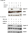
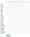
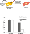
HIV-1 is dependent upon cellular co-factors to mediate its replication cycle in CD4(+) T cells and macrophages, the two major cell types infected by the virus in vivo. One critical co-factor is Cyclin T1, a subunit of a general RNA polymerase II elongation factor known as P-TEFb. Cyclin T1 is targeted directly by the viral Tat protein to activate proviral transcription. Cyclin T1 is up-regulated when resting CD4(+) T cells are activated and during macrophage differentiation or activation, conditions that are also necessary for high levels of HIV-1 replication. Because Cyclin T1 is a subunit of a transcription factor, the up-regulation of Cyclin T1 in these cells results in the induction of cellular genes, some of which might be HIV-1 co-factors. Using shRNA depletions of Cyclin T1 and transcriptional profiling, we identified 54 cellular mRNAs that appear to be Cyclin T1-dependent for their induction in activated CD4(+) T Jurkat T cells and during differentiation and activation of MM6 cells, a human monocytic cell line. The promoters for these Cyclin T1-dependent genes (CTDGs) are over-represented in two transcription factor binding sites, SREBP1 and ARP1. Notably, 10 of these CTDGs have been reported to be involved in HIV-1 replication, a significant over-representation of such genes when compared to randomly generated lists of 54 genes (p value<0.00021). The results of siRNA depletion and dominant-negative protein experiments with two CTDGs identified here, CDK11 and Casein kinase 1 gamma 1, suggest that these genes are involved either directly or indirectly in HIV-1 replication. It is likely that the 54 CTDGs identified here include novel HIV-1 co-factors. The presence of CTDGs in the protein space that was available for HIV-1 to sample during its evolution and acquisition of Tat function may provide an explanation for why CTDGs are enriched in viral co-factors.
Conflict of interest statement
Figures


Similar articles
-
Regulation of TAK/P-TEFb in CD4+ T lymphocytes and macrophages.Curr HIV Res. 2003 Oct;1(4):395-404. doi: 10.2174/1570162033485159. Curr HIV Res. 2003. PMID: 15049426 Review.
-
Transient induction of cyclin T1 during human macrophage differentiation regulates human immunodeficiency virus type 1 Tat transactivation function.J Virol. 2002 Nov;76(21):10579-87. doi: 10.1128/jvi.76.21.10579-10587.2002. J Virol. 2002. PMID: 12368300 Free PMC article.
-
Induction of the HIV-1 Tat co-factor cyclin T1 during monocyte differentiation is required for the regulated expression of a large portion of cellular mRNAs.Retrovirology. 2006 Jun 9;3:32. doi: 10.1186/1742-4690-3-32. Retrovirology. 2006. PMID: 16764723 Free PMC article.
-
Tat-associated kinase, TAK, activity is regulated by distinct mechanisms in peripheral blood lymphocytes and promonocytic cell lines.J Virol. 1998 Dec;72(12):9881-8. doi: 10.1128/JVI.72.12.9881-9888.1998. J Virol. 1998. PMID: 9811724 Free PMC article.
-
Regulatory functions of Cdk9 and of cyclin T1 in HIV tat transactivation pathway gene expression.J Cell Biochem. 1999 Dec 1;75(3):357-68. J Cell Biochem. 1999. PMID: 10536359 Review.
Cited by
-
Regulation of cyclin T1 and HIV-1 Replication by microRNAs in resting CD4+ T lymphocytes.J Virol. 2012 Mar;86(6):3244-52. doi: 10.1128/JVI.05065-11. Epub 2011 Dec 28. J Virol. 2012. PMID: 22205749 Free PMC article.
-
Transmembrane protein 106A is silenced by promoter region hypermethylation and suppresses gastric cancer growth by inducing apoptosis.J Cell Mol Med. 2014 Aug;18(8):1655-66. doi: 10.1111/jcmm.12352. Epub 2014 Jun 28. J Cell Mol Med. 2014. PMID: 24975047 Free PMC article.
-
Euphorbia Kansui Reactivates Latent HIV.PLoS One. 2016 Dec 15;11(12):e0168027. doi: 10.1371/journal.pone.0168027. eCollection 2016. PLoS One. 2016. PMID: 27977742 Free PMC article.
-
Control of HIV latency by epigenetic and non-epigenetic mechanisms.Curr HIV Res. 2011 Dec 1;9(8):554-67. doi: 10.2174/157016211798998736. Curr HIV Res. 2011. PMID: 22211660 Free PMC article.
-
Gene expression gradients along the tonotopic axis of the chicken auditory epithelium.J Assoc Res Otolaryngol. 2011 Aug;12(4):423-35. doi: 10.1007/s10162-011-0259-2. Epub 2011 Mar 12. J Assoc Res Otolaryngol. 2011. PMID: 21399991 Free PMC article.
References
-
- Zack JA, Arrigo SJ, Weitsman SR, Go AS, Haislip A, Chen IS. HIV-1 entry into quiescent primary lymphocytes: molecular analysis reveals a labile, latent viral structure. Cell. 1990;61:213–222. - PubMed
Publication types
MeSH terms
Substances
Grants and funding
LinkOut - more resources
Full Text Sources
Other Literature Sources
Molecular Biology Databases
Research Materials

