. 2008 Sep 5;3(9):e3153.
doi: 10.1371/journal.pone.0003153.

Targeted destruction of photosensitive retinal ganglion cells with a saporin conjugate alters the effects of light on mouse circadian rhythms

Affiliations

Targeted destruction of photosensitive retinal ganglion cells with a saporin conjugate alters the effects of light on mouse circadian rhythms

Didem Göz et al. PLoS One. .

Abstract

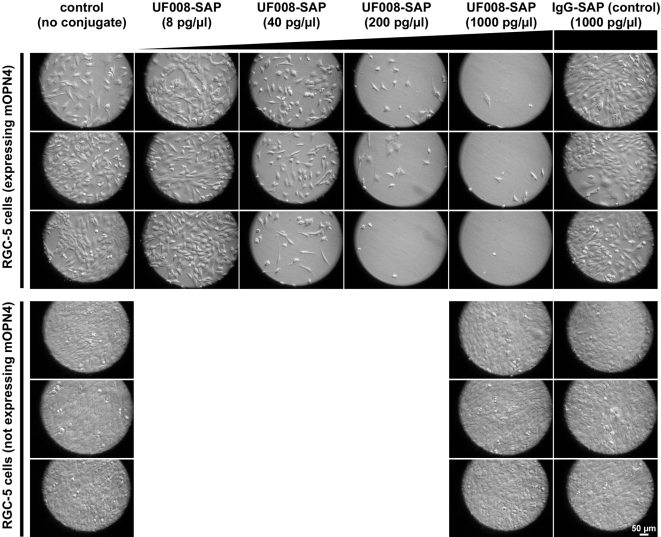
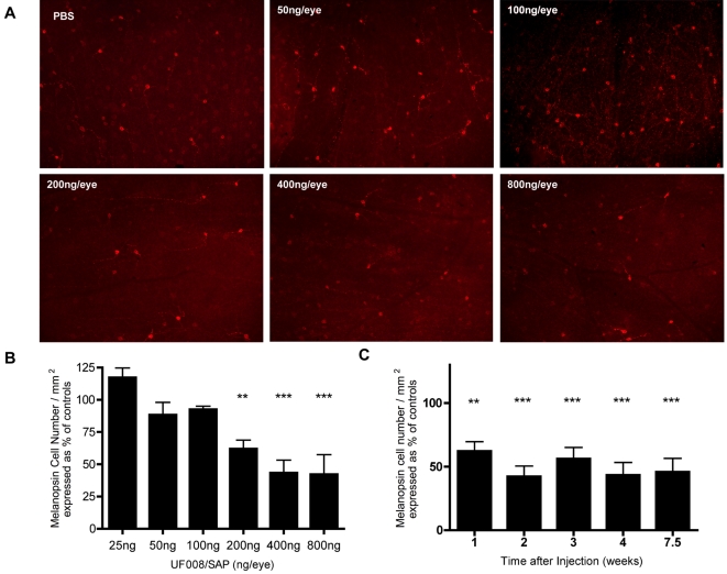
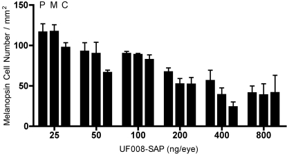
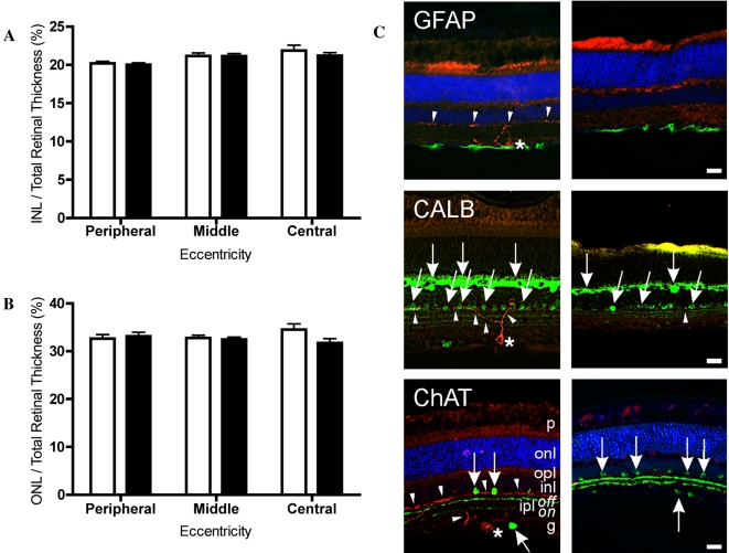
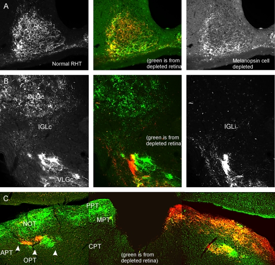
Non-image related responses to light, such as the synchronization of circadian rhythms to the day/night cycle, are mediated by classical rod/cone photoreceptors and by a small subset of retinal ganglion cells that are intrinsically photosensitive, expressing the photopigment, melanopsin. This raises the possibility that the melanopsin cells may be serving as a conduit for photic information detected by the rods and/or cones. To test this idea, we developed a specific immunotoxin consisting of an anti-melanopsin antibody conjugated to the ribosome-inactivating protein, saporin. Intravitreal injection of this immunotoxin results in targeted destruction of melanopsin cells. We find that the specific loss of these cells in the adult mouse retina alters the effects of light on circadian rhythms. In particular, the photosensitivity of the circadian system is significantly attenuated. A subset of animals becomes non-responsive to the light/dark cycle, a characteristic previously observed in mice lacking rods, cones, and functional melanopsin cells. Mice lacking melanopsin cells are also unable to show light induced negative masking, a phenomenon known to be mediated by such cells, but both visual cliff and light/dark preference responses are normal. These data suggest that cells containing melanopsin do indeed function as a conduit for rod and/or cone information for certain non-image forming visual responses. Furthermore, we have developed a technique to specifically ablate melanopsin cells in the fully developed adult retina. This approach can be applied to any species subject to the existence of appropriate anti-melanopsin antibodies.


Conflict of interest statement

Competing Interests: The authors have declared that no competing interests exist.

Figures

Figure 1
Figure 1. A saporin/anti-melanopsin(UF008) conjugate destroys cultured RGC-5 cells in a dose-dependent manner.
Cultures of RGC-5 cells, either stably expressing or not expressing mouse melanopsin, were exposed to a saporin conjugate (UF008/SAP) for 4 days. The concentrations of conjugates are shown above each column and the experiments were done in triplicate. Each panel represents a randomly selected field from a single well.
Figure 2
Figure 2. In vivo injection of UF008/SAP rapidly destroys ipRGCs in a dose-dependent manner.
UF008/SAP-dependent killing of ipRGCs in adult C57BL/6J mouse retina. A) Selected retinal flat-mount images (10× magnification) of PBS and UF008/SAP injected eyes from each dose group. B) Left eyes of mice (n = 5 per dose group) were injected with increasing doses of the UF008/SAP conjugate (25, 50, 100, 200, 400 and 800 ng/eye). Right eyes were injected with PBS to serve as controls. (1-way ANOVA with Tukey's Multiple Comparison Test, ** p<0.01, *** p<0.001). C) Mice (n = 4 per time group) were bilaterally injected with 400 ng of UF008/SAP conjugate per eye or 400 ng/eye of IgG/SAP (saporin conjugated to a “nonsense” rabbit IgG). At one week post-injection, the remaining number of melanopsin cells in the UF008/SAP injected retinas was significantly less than the controls, but the maximum melanopsin cell death was achieved at 2 weeks post-injection (1-way ANOVA with Tukey's Multiple Comparison Test, * p<0.05, *** p<0.001). The results for each experimental group in B and C were normalized relative to the data from the contralateral control eyes.
Figure 3
Figure 3. Destruction of ipRGCs by UF008/SAP depends on dose, but not on location within the retina.
Ablation of ipRGCs by UF008/SAP is dose-dependent, but not related to retinal eccentricity. Each triplet of bars represents data from “peripheral” (P), ”middle” (M), and “central” (C) fields, respectively. (2-way ANOVA with Bonferroni post-tests).
Figure 4
Figure 4. UF008/SAP kills ipRGCs without apparent damage to general retinal morphology.
UF008/SAP does not induce changes in gross retinal morphology. Eyes injected with 800 ng/eye of UF008/SAP conjugate did not differ ( 2-way ANOVA with Bonferroni post-test) from the PBS-injected control eyes in their relative (A) ONL or (B) INL thickness. (open bars, control eyes; black bars, UF008/SAP-injected). C) Cryostat sections through control retina (left column) and through UF008/SAP treated retina (right column). Red label (all images) - Melanopsin immunoreactive (IR) cells (*) and processes (arrowheads); Green label (Upper Row) - GFAP-IR glia at the base of the ganglion cell layer; Green label (Middle Row) - CALB-IR cells (arrows) in the outer and inner nuclear layers; Green label (Bottom row) - ChAT-IR amacrine cells (arrows) in the inner nuclear and ganglion cell layers between which are the two ChAT-IR terminal zones of the inner plexiform layer. Blue label - The nuclear stain, DAPI, most clearly reveals cells in the outer nuclear layer of the upper and lower images. Note the absence of melanopsin-IR staining in treated retinas (right column). Abbreviations of retinal layers: g – ganglion cell; inl – inner nuclear; ipl – inner plexiform (on and off sublayers); onl – outer nuclear; opl – outer plexiform; p – photoreceptor. Bar = 20 µm.
Figure 5
Figure 5. UF008/SAP greatly reduces retinal projections to the suprachiasmatic nucleus and intergeniculate leaflet.
CT-B tracing of RHT projections remaining after intravitreal UF008/SAP injection shows a terminal field densest along the lateral, ventrolateral and ventral SCN border, but greatly reduced in, or absent from, the dorso-central SCN. B) In the IGL, the contralateral projection from the UF008/SAP-injected retina is largely absent. C) There are no readily apparent differences with respect to the remaining retinal innervation of the OPT. The red label in all parts of the figure identifies terminals from the contralateral, undamaged retina. Abbreviations: APT – anterior pretectal n.; CPT – commissural pretectal n.; DLGc – dorsal lateral geniculate n;, contralateral; IGLc – intergeniculate leaflet, contralateral; IGLi – intergeniculate leaflet, ipsilateral; MPT – medial pretectal n.; NOT – nucleus of the optic tract; PPT – posterior pretectal n.; RHT – retinohypothalamic tract; VLGc – ventral lateral geniculate n., contralateral. (Ipsi- and contra- are referenced with respect to the injected with UF008/SAP).
Figure 6
Figure 6. UF008/SAP treatment has little effect on visual cliff performance.
Both UF008/SAP and IgG/SAP injected groups perform quite accurately in the visual cliff test, but there is a small deficit in the UF008/SAP treated group (p<0.05, Mann-Whitney test).
Figure 7
Figure 7. Depletion of ipRGCs greatly alters entrainment, period lengthening in LL and masking by LL.
Running records of a control and 3 UF008/SAP-treated mice showing phase angle of entrainment adopted in response to the gradual offset photoperiod, period during DD, during LL and re-entrainment (animals #139, 135) or failure to re-entrain (animals #122, 133) to LD12∶12 (grayed area indicates darkness). The phase angle of entrainment (Φ) during the gradual offset photoperiod is indicated for each individual (ZT12 = light completely off). B) Daily irradiance pattern recorded with a Gigahertz-Optik P-9710-2 universal optometer measured at cage level during the gradual offset light-dark paradigm. C) Relationship between remaining ipRGC density and the stable phase angle of entrainment of adopted by UF008/SAP injected mice and controls during the gradual offset photoperiod. Note the cluster of controls and the outlier animal (green-filled circle), which was injected with the saporin conjugate, but has not lost its ipRGCs. A second outlier animal (red-filled circle) had a normal phase angle of entrainment despite greatly reduced ipRGC density. D) There was no difference in period in circadian period during DD between UF008/SAP and IgG/SAP injected mice, but in LL, IgG/SAP injected mice significantly lengthened their periods (p<0.001; paired t test), becoming significantly different from UF008/SAP injected animals (p<.001, unpaired t test) which did not show any period lengthening in response to LL. E) LL induced masking was absent in UF008/SAP injected mice. Revolutions per day for the last 5 days in DD were compared to those during the initial 5 days of LL. For UF008/SAP mice, revolutions during DD and LL did not differ; for controls, revolutions/24 hr dropped by about 40% (p<.001; paired t tests).
Figure 8
Figure 8. Masking response to a light pulse is absent in mice that lose entrainment to LD12∶12.
Masking is nearly normal by UF008/SAP injected mice in response to a 1 hr light pulse. However, the animals recovered more slowly than controls. There is no effect of treatment or of time, but there is a significant interaction (1 way ANOVA with repeated measures, p<.004). In mice that lost entrainment subsequent to UF008/SAP treatment, a 1 hr light pulse failed to induce masking.

Similar articles

Cited by

References

    1. Berson DM, Dunn FA, Takao M. Phototransduction by retinal ganglion cells that set the circadian clock. Science. 2002;295:1070–1073. - PubMed
    1. Gooley JJ, Lu J, Chou TC, Scammell TE, Saper CB. Melanopsin in cells of origin of the retinohypothalamic tract. Nat Neurosci. 2001;4:1165. - PubMed
    1. Hattar S, Kumar M, Park A, Tong P, Tung J, et al. Central projections of melanopsin-expressing retinal ganglion cells in the mouse. J Comp Neurol. 2006;497:326–349. - PMC - PubMed
    1. Yoshimura T, Nishio M, Goto M, Ebihara S. Differences in circadian photosensitivity between retinally degenerate CBA/J mice ( rd/rd ) and normal CBA/N mice (+/+). Journal of Biological Rhythms. 1994;9:51–60. - PubMed
    1. Foster RG, Provencio I, Hudson D, Fiske S, De Grip W, et al. Circadian photoreception in the retinally degenerate mouse (rd/rd). J Comp Physiol [A] 1991;169:39–50. - PubMed

Publication types

MeSH terms