. 2008 Sep 5;3(9):e3145.
doi: 10.1371/journal.pone.0003145.

Neuroprotective effect of transplanted human embryonic stem cell-derived neural precursors in an animal model of multiple sclerosis

Affiliations

Neuroprotective effect of transplanted human embryonic stem cell-derived neural precursors in an animal model of multiple sclerosis

Michal Aharonowiz et al. PLoS One. .

Abstract

Background: Multiple sclerosis (MS) is an immune mediated demyelinating disease of the central nervous system (CNS). A potential new therapeutic approach for MS is cell transplantation which may promote remyelination and suppress the inflammatory process.

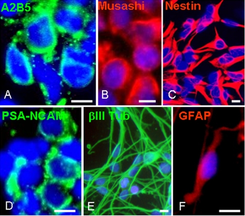
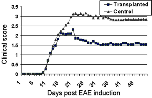
Methods: We transplanted human embryonic stem cells (hESC)-derived early multipotent neural precursors (NPs) into the brain ventricles of mice induced with experimental autoimmune encephalomyelitis (EAE), the animal model of MS. We studied the effect of the transplanted NPs on the functional and pathological manifestations of the disease.

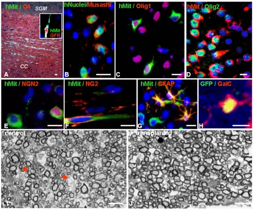
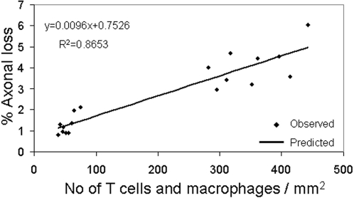
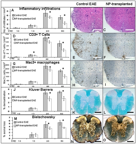
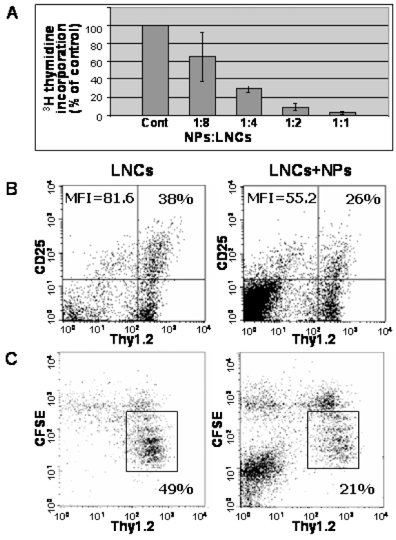
Results: Transplanted hESC-derived NPs significantly reduced the clinical signs of EAE. Histological examination showed migration of the transplanted NPs to the host white matter, however, differentiation to mature oligodendrocytes and remyelination were negligible. Time course analysis of the evolution and progression of CNS inflammation and tissue injury showed an attenuation of the inflammatory process in transplanted animals, which was correlated with the reduction of both axonal damage and demyelination. Co-culture experiments showed that hESC-derived NPs inhibited the activation and proliferation of lymph node-derived T cells in response to nonspecific polyclonal stimuli.

Conclusions: The therapeutic effect of transplantation was not related to graft or host remyelination but was mediated by an immunosuppressive neuroprotective mechanism. The attenuation of EAE by hESC-derived NPs, demonstrated here, may serve as the first step towards further developments of hESC for cell therapy in MS.


Conflict of interest statement

Competing Interests: BR holds shares and is the chief scientific officer of CellCure Neurosciences Ltd. TBH also holds shares and is a consultant of CellCure Neurosciences. CellCure neurosciences develops hESC-derived neural progeny for cell therapy for neurological disorders.

Figures

Figure 1
Figure 1. Characterization of the hESC-derived spheres prior to transplantation.
Spheres enriched for NPs were developed from hESCs that were cultured for 3 weeks as free floating clusters in serum-free medium supplemented with BFGF, EGF and noggin. The spheres were further expanded for 4–6 weeks in the same medium supplemented with bFGF and EGF before transplantation. For in-vitro characterization spheres were triturated and plated on fibronectin-coated coverslips. Immunofluorescent stainings, 2 hours after plating, demonstrated that the majority of the cells within the spheres were immunoreactive with anti-A2B5 (A), -Musashi (B), -Nestin (C) and -PSA-NCAM (D). To induce differentiation, the plated cells were further cultured 7 days in the absence of mitogens. Immunofluorescent stainings showed that the NPs differentiated mainly into βIII tubulin-expressing neurons (E) and GFAP expressing astrocytes (F). Cells expressing oligodendroglial markers were not observed. Nuclei in A–F are counterstained with DAPI (blue). Scale bars: 15 µm.
Figure 2
Figure 2. The clinical course of EAE is attenuated after transplantation of hESC-derived NPs.
hESC-derived NPs were transplanted into the lateral brain ventricles of EAE mice and the severity of clinical signs was scored daily. A significant attenuation of the clinical score was observed in NPs-transplanted animals (▪) in comparison to vehicle-transplanted control animals (▴). The reduced severity of clinical scores in NP-transplanted animals was evident as early as the acute phase of the disease (day 19 post-EAE induction). See material and methods section for details regarding the clinical scoring system.
Figure 3
Figure 3. Transplanted NPs migrate into white matter tracts of EAE brains, differentiate into neural progenitors and do not promote remyelination.
(A–H) Immunofluorescence stainings of brain sections demonstrating the migration and differentiation of transplanted NPs, which were identified by the expression of human mitochondria (A, C–G), human nuclear antigens (B) and GFP (inset in A, H). (A): The NPs migrated extensively into white matter areas of the CNS such as the corpus callosum (CC) and were not observed in grey areas such as subcortical grey matter (SGM). Co-staining against the oligodendroglial marker, O4 (red) was used to identify the white matter. (B–H): Most of the transplanted cells either remained as uncommitted NPs expressing Musashi (B) or differentiated into early neuronal/oligodendroglial progenitors expressing Olig1 (C) or Olig2 (D). Further differentiation into more committed neuronal progenitors, oligodendrocyte progenitors or astrocytes expressing NGN2 (E), NG2 (F) and GFAP (G), respectively, was infrequent (∼1% of the transplanted cells per each of the cell types). Terminal differentiation of the transplanted NPs into GalC-expressing mature oligodendrocytes (H) was rare (<0.01% of the transplanted cells). Nuclei in A–H are counterstained with DAPI (blue). I–J: Toluidine blue stained transverse semi-thin sections of resin embedded spinal cords of NP-transplanted (J) and control (I) animals. In NP-transplanted animals, there were less demyelinated (arrows in I) and more normally myelinated axons than in controls. In both groups there were very rare remyelinated axons (G ratio>0.8). Scale bars: 15 µm (A–H), 5 µm (I–J).
Figure 4
Figure 4. The extent of the inflammatory process strongly correlates with the severity of tissue damage in the acute phase of EAE.
NP- transplanted and control mice were sacrificed at 10, 13, 20 and 50 days post EAE induction for pathological analysis of inflammation and tissue damage. Linear-regression analysis of the numbers of inflammatory cells and the extent of axonal loss in both NP-transplanted and control EAE mice at 13 and 20 days post EAE induction showed a strong correlation between the numbers of T cells and macrophages per mm2 and the percentage of axonal loss.
Figure 5
Figure 5. Attenuation of the progression of inflammation and tissue damage in the CNS of NPs-transplanted mice.
Pathological examination of spinal cord sections from NP-transplanted and control mice were performed at 10, 13, 20 and 50 days post EAE induction to evaluate CNS inflammation, demyelination and axonal damage. In NP-transplanted mice an attenuation of the number of immune-cell infiltrates (A), T cells (D) and macrophages/activated microglia (G) per mm2 was evident from day 13 post EAE induction and became significant at days 20 and 50. Demyelination and axonal damage, which were analyzed by loss of Kluver Barrera (J), and Bielschowsky staining (M), respectively, were both significantly reduced at day 20 and 50 post EAE induction. The differences between the study and control groups in the severity of all parameters gradually increased with time. Representative day 20 images of H&E staining (B, C), immunostaining for CD3 (E, F) and Mac3 (H, I), Kluver Barrera staining (K, L) and Bielschowsky silver staining (N, O). * P<0.05. Scale bars: 100 µm.
Figure 6
Figure 6. Direct suppressive effects of hESC-derived NPs on lymph node cells (LNCs) and T cells derived from naïve C57BL mice.
LNCs derived from naïve mice were co-cultured with NPs and activated with ConA. The NPs suppressed 3H-thymidine incorporation into the activated LNCs in a dose-dependent manner (A). FACS analysis of interleukin-2 receptor α (IL-2Rα; CD25) expression after 24 hours of ConA-stimulation showed that human NPs inhibited the activation of Thy1.2+ T cells as determined by the fraction of labeled cells and by mean fluorescent intensity (MFI) (B). FACS analysis of CFSE labeled LNCs after 72 hours of ConA stimulation showed that co-culturing with the human NPs inhibited the proliferation of Thy1.2+ T cells. The fraction of T cells that have proliferated and therefore diluted the CFSE fluorescence (within the rectangle) was reduced in the presence of the human NPs (C).

References

    1. Gironi M, Bergami A, Brambilla E, Ruffini F, Furlan R, et al. Immunological markers in multiple sclerosis. Neurol Sci. 2000;21(4 Suppl 2):S871–875. - PubMed
    1. Hemmer B, Cepok S, Nessler S, Sommer N. Pathogenesis of multiple sclerosis: an update on immunology. Curr Opin Neurol. 2002;15:227–231. - PubMed
    1. Lassmann H. Mechanisms of demyelination and tissue destruction in multiple sclerosis. Clin Neurol Neurosurg. 2002;104:168–171. - PubMed
    1. Franklin RJ. Why does remyelination fail in multiple sclerosis? Nat Rev Neurosci. 2002;3:705–714. - PubMed
    1. Kornek B, Storch MK, Weissert R, Wallstroem E, Stefferl A, et al. Multiple sclerosis and chronic autoimmune encephalomyelitis: a comparative quantitative study of axonal injury in active, inactive, and remyelinated lesions. Am J Pathol. 2000;157:267–276. - PMC - PubMed

Publication types

Substances