. 2008 Sep 5;4(9):e1000170.
doi: 10.1371/journal.pgen.1000170.

Heterokaryon-based reprogramming of human B lymphocytes for pluripotency requires Oct4 but not Sox2

Affiliations

Heterokaryon-based reprogramming of human B lymphocytes for pluripotency requires Oct4 but not Sox2

Carlos F Pereira et al. PLoS Genet. .

Abstract

Differentiated cells can be reprogrammed through the formation of heterokaryons and hybrid cells when fused with embryonic stem (ES) cells. Here, we provide evidence that conversion of human B-lymphocytes towards a multipotent state is initiated much more rapidly than previously thought, occurring in transient heterokaryons before nuclear fusion and cell division. Interestingly, reprogramming of human lymphocytes by mouse ES cells elicits the expression of a human ES-specific gene profile, in which markers of human ES cells are expressed (hSSEA4, hFGF receptors and ligands), but markers that are specific to mouse ES cells are not (e.g., Bmp4 and LIF receptor). Using genetically engineered mouse ES cells, we demonstrate that successful reprogramming of human lymphocytes is independent of Sox2, a factor thought to be required for induced pluripotent stem (iPS) cells. In contrast, there is a distinct requirement for Oct4 in the establishment but not the maintenance of the reprogrammed state. Experimental heterokaryons, therefore, offer a powerful approach to trace the contribution of individual factors to the reprogramming of human somatic cells towards a multipotent state.


Conflict of interest statement

The authors have declared that no competing interests exist.

Figures
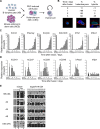
Figure 1
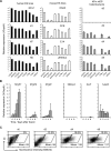
Figure 1. Pluripotent reprogramming of human B-lymphocytes by mouse ES cells is initiated in heterokaryons prior to nuclear fusion and cell division.
(A) Shows the experimental strategy used to generate interspecies heterokaryons (hB x mES). Human B-lymphocytes (hB) and mouse embryonic stem cells (mES) were respectively labelled with the cell membrane dyes DiI and DiD and fused in the presence of polyethylene glycol (PEG). Fused cells were FACS sorted and cultured under conditions that promote mES self-renewal. (B) Immunofluorescence analysis of the kinetics of heterokaryon (cells in which parental nuclei share the same cytoplasm but remain discrete) and hybrid formation (where both parental genomes occupy the same nucleus) following PEG induced fusion. In the lower panels hB-derived nuclei were distinguished by mouse nuclei on basis of DAPI (blue) and human Lamin A/C staining (green), and actin staining (red) delineates individual cells. Confocal sections showing a hB cell prior to fusion (left, day 0), a heterokaryon [one mouse (with DAPI intense foci) and one human nucleus (hLamin A/C positive)] (middle, day 2) and a hybrid cell (right, day 3) are shown. Scale bar, 10 µm. n = 100. (C) The expression of hES-specific genes (hOct4, hNanog, hCripto, hDnmt3b, hTle1, hRex1) was assessed by quantitative RT-PCR analysis 0 to 8 days after cell fusion. Positive (hES-NCL1, black bars) and negative (hB) controls for this analysis were included. (D) Activation of embryonic genes is accompanied by silencing of lymphocyte-specific genes (hCD19, hCD37, hCD20, hCD45 and hPax5), while a constitutively expressed gene hHprt remained detectable at similar levels at all time points. Data were normalised to hGapdh expression. Error bars indicate the s.d. of 3 independent experiments. (E) Bisulfite genomic sequencing analysis of DNA methylation at the human Oct4 promoter 0, 2, 4 and 8 days after cell fusion demonstrated the rapid de-methylation of Oct4 induced by fusion with mES cells. Human ES cells (hES, cell line H1; lower panel) are shown as controls. The methylation pattern of Igf2/H19 imprinting control region (ICR) remained unaltered throughout the experiment. The position of CpG sites relative to the transcriptional start site (TSS) is indicated. Open circles represent unmethylated cytosines, black closed circles represent methylated cytosines and grey closed circles represent constitutively methylated cytosines. Regions 1, 2 and 3 indicate CpG sites that are part of the same PCR product.
Figure 2
Figure 2. Gene expression in reprogrammed lymphocytes resembles human rather than mouse ES cell lines.
(A) Quantitative RT-PCR analysis of the relative levels of gene expression in several human (NCL1, H1, H7 and H9), mouse (OS25, CCE, E14 and ZHBTc4) ES cell lines and in reprogrammed lymphocytes (hB x mES) at 0, 2, 4 and 8 days after cell fusion. Human ES lines (left panel) and hB x mES (right panel) gene expression data were normalised to hGapdh. Mouse ES lines (middle panel) gene expression data were normalised to mGapdh. Error bars indicate the s.d. of 3–4 independent experiments. (B) After cell fusion, genes involved in the maintenance of undifferentiated human ES cells (hFgfr1, hFgfr2 and hFgf2) were activated while genes selectively expressed by mouse ES cells (hBmp4, hLifr and hJak3) were not induced. Data were normalised to hGapdh expression. Error bars indicate the s.d. of 3 independent experiments. (C) Heterokaryons resulting from human B cell and mouse ES cell fusions (hB x mES) were stained for SSEA4 at 0, 2, 4 and 8 days and expression was analysed by flow cytometry. The results showed that 13.5% (day 2), 16.6% (day 4) and 15.8% (day 8) of total heterokaryons expressed SSEA4, as delineated by the rhomboid gates. Mean intensity fluorescence of positive cells is indicated.
Figure 3
Figure 3. Multi-lineage potential is reset in reprogrammed human lymphocytes.
(A) Hybrid colonies resulting from fusion of human B cells (hB) and mouse ES cells (hB x mES) showed alkaline phosphatase activity (pink), that was reduced upon retinoic acid (RA) treatment. (B) RA treatment of hybrid colonies (day 6) generated cells that expressed Nestin (green) detected by immunostaining using an antibody specific for human (and not mouse) Nestin protein. DAPI counterstaining (blue) is shown. Scale bars, 50 µm. (C) Quantitative RT-PCR analysis of gene expression upon RA treatment of hybrid cells (blue line) showed that levels of pluripotency genes (hOct4, hNanog and hRex1) declined while differentiation-associated genes were upregulated [extra-embryonic (hCdx2, hHand1 and hGata6), endoderm (hSox7, hHnf4 and hCollagenIVαI), mesoderm (hMixl1, hEbf and hMyoD) and ectoderm (hNestin)]. Unfused hB cells were included as controls (black line). Data were normalised to hGapdh expression. (D) Bisulfite genomic sequencing analysis of DNA methylation at the human Oct4 promoter 8 days after RA treatment showed the re-methylation of the Oct4 promoter while the Igf2/H19 imprinted control region (ICR) remains unaltered. The position of CpG sites relative to the transcriptional start site (TSS) is indicated. Open circles represent unmethylated cytosines, black closed circles represent methylated cytosines and grey closed circles represent constitutively methylated cytosines.
Figure 4
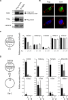
Figure 4. Oct4 is required for successful reprogramming.
(A) Mouse ES cells expressing a tagged Oct4 protein (Flag-mOct4) were generated by insertion of Flag-tagged mouse Oct4 cDNA in E14tg2a ES cells (parental cell line). Western blotting with anti-Oct4 and anti-Flag antibodies confirmed the presence of Flag-tagged Oct4 protein by transduced cells. Equivalent protein loading is shown with Lamin B detection. (B) Immunofluorescence analysis of cultured heterokaryons 6 hours after cell fusion showed the presence of ES cell-derived Oct4 (Flag-Oct4, green) in a human nucleus (arrowed). Human nuclei were distinguished from mouse nuclei on basis of diffuse versus punctuate DAPI staining (blue), respectively. Actin labelling (red) delineates the cell membrane. Images are confocal sections of heterokaryons containing a single mouse (with DAPI intense foci) and a single human nucleus. Scale bar, 10 µm. (C) In ZHBTc4 ES cells endogenous Oct4 was replaced by an inducible transgene (Oct4βgeo) which can be downregulated by addition of doxycycline (Dox) . Quantitative RT-PCR analysis showed that 6 hours (+6) and 12 hours (+12) after Dox treatment, mOct4 was progressively downregulated, while expression of other pluripotency-associated genes (mNanog, mCripto, mRex1 and mSox2) was largely unaffected. (D) ES cells expressing normal levels of Oct4 (-), partially reduced (Dox+6) or lacking Oct4 expression (Dox+12) were fused to hB-lymphocytes. Successful reprogramming was assessed by quantifying the abundance of human ES-associated transcripts two days after fusion by qRT-PCR. Activation of pluripotency genes in hB-lymphocytes was reduced or impaired when Oct4 was ablated. Data were normalised to Gapdh expression. Error bars indicate the s.d. of 2–3 independent experiments.
Figure 5
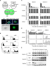
Figure 5. Sox2 is dispensable for reprogramming.
(A) In 2TS22C ES cells endogenous Sox2 is replaced by an inducible transgene (Sox2Zeo) which can be downregulated by addition of doxycycline (Dox) . Quantitative RT-PCR analysis showed that 12 hours (+12) and 24 hours (+24) after Dox treatment, mSox2 was downregulated while expression of other pluripotency-associated genes (mNanog, mCripto, mRex1 and mOct4) continued to be expressed. 2O1 ES cells are Sox2-deficient mES cells (asterisk) in which mOct4 expression is up-regulated (red bars). (B) ES cells expressing Sox2 (-), Sox2 depleted cells (Dox+12, Dox+24) and 2O1 cells were fused to hB-lymphocytes. Successful reprogramming was assessed by quantifying the abundance of human ES-associated transcripts two days after fusion by qRT-PCR. Activation of pluripotency genes in hB-lymphocytes occurs in the absence of mSox2. An elevated induction of hSox2 using 2O1 cells as a fusion partner is highlighted by an arrow (red). All data were normalised to Gapdh expression and error bars indicate the s.d. of 2–3 independent experiments.
Figure 6
Figure 6. Reprogramming is self-sustaining and can be maintained in the absence of ES-derived Oct4.
(A) To address whether reprogramming is stable or subject to reversion, we ablated Oct4 expression after hybrid formation. ZHBTc4 ES cells [mES with endogenous Oct4 replaced by an inducible transgene (Oct4βgeo) which can be downregulated by addition of doxycycline (Dox)] were fused with mouse B-lymphocytes (mB) carrying a GFP transgene under the control of Oct4 promoter (GOF18ΔPE). Reprogramming of mB results in the re-activation of GFP in hybrid colonies (d10, lower panels). Kinetic analysis of single cells (upper panels) showed that transgene re-activation occurs in heterokaryons (day 2, 2 arrows), and hybrid cells (day3, arrowhead). mB cells are shown as negative controls. Nuclei were visualised with DAPI staining (blue). Scale bars, 10 µm. (B) Hybrid clones (mES x mB, 4n) that re-expressed GFP were isolated and analysed by FACS. mES, mB and mES x mB hybrid cells unstained (left panel) or stained with propidium iodide (right panel) to assess GFP expression and DNA content, respectively. (C) Hybrid clones (4 and 12) were treated with Dox to ablate ES-derived Oct4, and quantitative RT-PCR confirmed downregulation of Oct4βgeo transcript (upper panels). Removal of mES-derived Oct4βgeo in hybrid clones did not affect gene expression of pluripotency-associated transcripts (lower panels; mOct4, mNanog and mSox2) after 96 hours of Dox treatment. (D) No differentiation was observed after Oct4 removal in hybrid cells. mRex1 expression was retained and the extra-embryonic markers mHand1 and mCdx2 were not induced. In ZHBTc4 ES cells (open bars) upon Dox treatment, mHand1 and mCdx2 were induced and are shown for comparison. Data were normalised to mGapdh expression. Error bars indicate the s.d. of 3 independent experiments. (E) Western blotting with anti-Oct4 antibody confirmed that Oct4 protein is rapidly removed after Dox treatment of ZHBTc4 ES cells (lower panel) but remains detectable at all times in hybrid cells (upper panels). Equivalent protein loading is shown with Lamin B detection.

Similar articles

Cited by

References

    1. Hochedlinger K, Jaenisch R. Monoclonal mice generated by nuclear transfer from mature B and T donor cells. Nature. 2002;415:1035–1038. - PubMed
    1. Wilmut I, Schnieke AE, McWhir J, Kind AJ, Campbell KH. Viable offspring derived from fetal and adult mammalian cells. Nature. 1997;385:810–813. - PubMed
    1. Gurdon JB. Adult frogs derived from the nuclei of single somatic cells. Dev Biol. 1962;4:256–273. - PubMed
    1. Byrne JA, Pedersen DA, Clepper LL, Nelson M, Sanger WG, et al. Producing primate embryonic stem cells by somatic cell nuclear transfer. Nature. 2007;450:497–502. - PubMed
    1. Davis RL, Weintraub H, Lassar AB. Expression of a single transfected cDNA converts fibroblasts to myoblasts. Cell. 1987;51:987–1000. - PubMed

Publication types

MeSH terms