

Segmental duplications arise from Pol32-dependent repair of broken forks through two alternative replication-based mechanisms
- PMID: 18773114
- PMCID: PMC2518615
- DOI: 10.1371/journal.pgen.1000175
Segmental duplications arise from Pol32-dependent repair of broken forks through two alternative replication-based mechanisms
Abstract
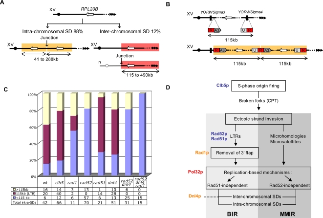
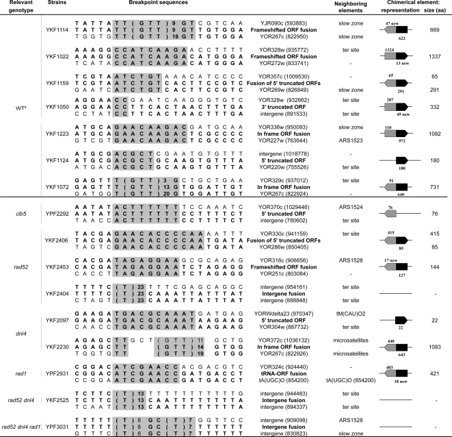
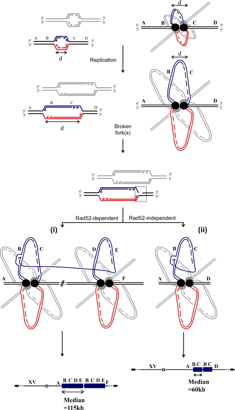
The propensity of segmental duplications (SDs) to promote genomic instability is of increasing interest since their involvement in numerous human genomic diseases and cancers was revealed. However, the mechanism(s) responsible for their appearance remain mostly speculative. Here, we show that in budding yeast, replication accidents, which are most likely transformed into broken forks, play a causal role in the formation of SDs. The Pol32 subunit of the major replicative polymerase Poldelta is required for all SD formation, demonstrating that SDs result from untimely DNA synthesis rather than from unequal crossing-over. Although Pol32 is known to be required for classical (Rad52-dependant) break-induced replication, only half of the SDs can be attributed to this mechanism. The remaining SDs are generated through a Rad52-independent mechanism of template switching between microsatellites or microhomologous sequences. This new mechanism, named microhomology/microsatellite-induced replication (MMIR), differs from all known DNA double-strand break repair pathways, as MMIR-mediated duplications still occur in the combined absence of homologous recombination, microhomology-mediated, and nonhomologous end joining machineries. The interplay between these two replication-based pathways explains important features of higher eukaryotic genomes, such as the strong, but not strict, association between SDs and transposable elements, as well as the frequent formation of oncogenic fusion genes generating protein innovations at SD junctions.
Conflict of interest statement
The authors have declared that no competing interests exist.
Figures
Similar articles
-
DNA REPAIR. Mus81 and converging forks limit the mutagenicity of replication fork breakage.Science. 2015 Aug 14;349(6249):742-7. doi: 10.1126/science.aaa8391. Science. 2015. PMID: 26273056 Free PMC article.
-
Break-induced replication and telomerase-independent telomere maintenance require Pol32.Nature. 2007 Aug 16;448(7155):820-3. doi: 10.1038/nature06047. Epub 2007 Aug 1. Nature. 2007. PMID: 17671506
-
Chromosomal translocations caused by either pol32-dependent or pol32-independent triparental break-induced replication.Mol Cell Biol. 2009 Oct;29(20):5441-54. doi: 10.1128/MCB.00256-09. Epub 2009 Aug 3. Mol Cell Biol. 2009. PMID: 19651902 Free PMC article.
-
MMEJ repair of double-strand breaks (director's cut): deleted sequences and alternative endings.Trends Genet. 2008 Nov;24(11):529-38. doi: 10.1016/j.tig.2008.08.007. Epub 2008 Sep 21. Trends Genet. 2008. PMID: 18809224 Free PMC article. Review.
-
Exploring the roles of Mus81-Eme1/Mms4 at perturbed replication forks.DNA Repair (Amst). 2007 Jul 1;6(7):1004-17. doi: 10.1016/j.dnarep.2007.02.019. Epub 2007 Apr 3. DNA Repair (Amst). 2007. PMID: 17409028 Review.
Cited by
-
Microhomology directs diverse DNA break repair pathways and chromosomal translocations.PLoS Genet. 2012;8(11):e1003026. doi: 10.1371/journal.pgen.1003026. Epub 2012 Nov 8. PLoS Genet. 2012. PMID: 23144625 Free PMC article.
-
Divergent roles for the two PolI-like organelle DNA polymerases of Arabidopsis.Plant Physiol. 2011 May;156(1):254-62. doi: 10.1104/pp.111.173849. Epub 2011 Mar 22. Plant Physiol. 2011. PMID: 21427281 Free PMC article.
-
Translesion Polymerases Drive Microhomology-Mediated Break-Induced Replication Leading to Complex Chromosomal Rearrangements.Mol Cell. 2015 Dec 17;60(6):860-72. doi: 10.1016/j.molcel.2015.10.041. Epub 2015 Dec 6. Mol Cell. 2015. PMID: 26669261 Free PMC article.
-
Pathways and Mechanisms that Prevent Genome Instability in Saccharomyces cerevisiae.Genetics. 2017 Jul;206(3):1187-1225. doi: 10.1534/genetics.112.145805. Genetics. 2017. PMID: 28684602 Free PMC article. Review.
-
POLD3 as Controller of Replicative DNA Repair.Int J Mol Sci. 2024 Nov 19;25(22):12417. doi: 10.3390/ijms252212417. Int J Mol Sci. 2024. PMID: 39596481 Free PMC article. Review.
References
-
- Bailey JA, Eichler EE. Primate segmental duplications: crucibles of evolution, diversity and disease. Nat Rev Genet. 2006;7:552–564. - PubMed
-
- Sharp AJ, Hansen S, Selzer RR, Cheng Z, Regan R, et al. Discovery of previously unidentified genomic disorders from the duplication architecture of the human genome. Nat Genet. 2006;38:1038–1042. - PubMed
-
- Courseaux A, Nahon JL. Birth of two chimeric genes in the Hominidae lineage. Science. 2001;291:1293–1297. - PubMed
Publication types
MeSH terms
Substances
LinkOut - more resources
Full Text Sources
Other Literature Sources
Molecular Biology Databases
Research Materials
