. 2009 Feb;238(2):358-66.
doi: 10.1002/dvdy.21681.

Fgf16(IRESCre) mice: a tool to inactivate genes expressed in inner ear cristae and spiral prominence epithelium

Affiliations

Fgf16(IRESCre) mice: a tool to inactivate genes expressed in inner ear cristae and spiral prominence epithelium

Ekaterina P Hatch et al. Dev Dyn. 2009 Feb.

Abstract

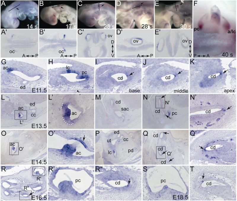
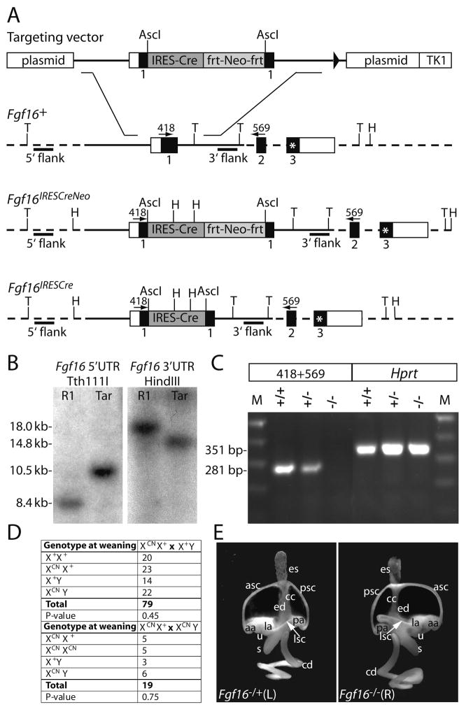
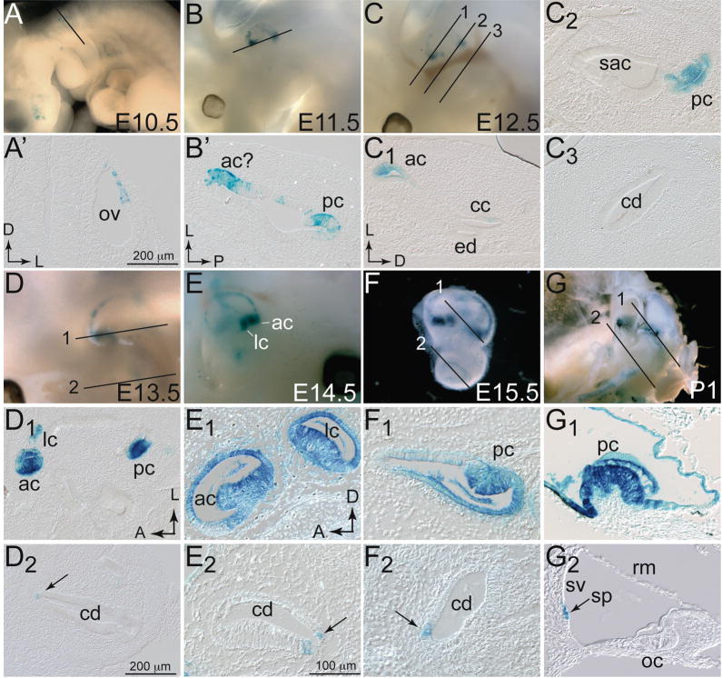
Fibroblast growth factors play important roles in inner ear development. Previous studies showed that mouse Fgf16 is expressed asymmetrically during the otic cup and vesicle stages of development, suggesting roles in regulating or responding to anteroposterior axial cues. Here, we studied otic Fgf16 expression throughout embryonic development and found transcripts in the developing cristae and in a few cells in the lateral wall of the cochlear duct. To determine the otic function of Fgf16 and to follow the fate of Fgf16-expressing cells, we generated an Fgf16(IRESCre) allele. We show that Fgf16 does not have a unique role in inner ear development and that the Fgf16 lineage is found throughout the three cristae, in portions of the semicircular canal ducts, and in the cochlear spiral prominence epithelial cells. This strain will be useful for gene ablations in these tissues.


Figures

Figure 1
Figure 1. Fgf16 mRNA expression from E9.0–E18.5
Whole-mount 14-somite (A), 17-somite (B), 23-somite (C), and 28-somite (D), 30-somite (E), and 40-somite (F) embryos were probed with labeled antisense Fgf16 cRNA. Anterior is to left (A,B,C,D,E) or right (F). White lines indicate the plane of coronal (A′,B′,D′) or transverse (D′,E′) sections shown in panels below the whole mounts, black arrows indicate the otic vesicle. Fgf16 expression in transverse (G-R″) or sagittal (S,T) paraffin sections. (L′) Solid white line indicates strong expression in anterior crista, dotted white line indicates weaker expression in cells adjacent to the crista. Abbreviations: A, anterior; ac, anterior crista; a/lc, anterior and lateral criate; cc, common crus; cd, cochlear duct; D, dorsal; ed, endolymphatic duct; lc, lateral crista; oc, otic cup; ov, otic vesicle; P, posterior: pc, posterior crista; sac, saccule; ut, utricle; V, ventral.
Figure 2
Figure 2. Gene targeting at the Fgf16 locus
(A) Structure of the linearized Fgf16 targeting vector and depiction of the wild type Fgf16 allele (Fgf16+), the correctly targeted mutant allele in ES cells and offspring of chimeric mice (Fgf16IRESCreNeo) and the targeted allele after exposure to FLP recombinase (Fgf16IRESCre). Mouse genomic Fgf16 DNA is depicted with solid lines; dotted lines indicate Fgf16 genomic DNA that is not present in the targeting vector; untranslated regions are shown as open boxes, protein coding regions as solid boxes. The IRES-Cre cassette is shown as a dark grey box; the Neo cassette flanked by frt sites as a light grey box; the stop codon in the Fgf16 frame in exon 3 as an asterisk. The plasmid backbone and the thymidine kinase (TK1) gene are depicted as open boxes. Recognition sites for Tth111I and HindIII, used in Southern analysis are indicated by “T” and “H” respectively. Probes used for Southern analysis are shown as black bars. Numbered arrows indicate the identity, position and directionality of primers used for the RT-PCR assay. (B) Southern blot hybridization assay demonstrating correct targeting of Fgf16 in ES cells. Tth111I-digested genomic DNA from either the R1–45 ES cell line (R1) or a correctly targeted cell line (Tar) probed with the external 5′ probe. (C) RT-PCR assay used to detect Fgf16 mRNA in wild type and mutant animals. Total E9.0 RNA isolated from female wild type (+/+), female heterozygous (+/−) and male hemizygous mutant (−/Y) embryos was PCR-amplified using primers 418 and 569. A 351 bp Hprt cDNA fragment was amplified from all samples as a positive control. (D) Fgf16 backcross and intercross genotypes. Offspring of the indicated crosses were genotyped at weaning. P-values were determined using the method of χ2. The Fgf16IRESCreNeo allele is defined as XNeo. (E) Fgf16-deficient inner ears were morphologically normal. Representative lateral views of paint-filled inner ears from E15.5 female heterozygous control and Fgf16IRESCreNeo homozygous mutants. Labeled structures: aa, anterior ampulla; asc, anterior semicircular canal; cc, common crus; cd, cochlear duct; ed, endolymphatic duct; es, endolymphatic sac; la, lateral ampulla; lsc, lateral semicircular canal; pa, posterior ampulla; psc, posterior semicircular canal; s, saccule; u, utricle.
Figure 3
Figure 3. Lineage of Fgf16-expressing cells at E10.5-P1
Embryos (A–D), inner ears (E,F) or heads (G) harboring both the Rosa26LacZ reporter and Fgf16CreNeo alleles were stained with X-gal at E10.5 (A), E11.5 (B), E12.5 (C), E13.5 (D), E14.5, (E) E15.5 (F), and P1 (G). Anterior is to left in all whole mount panels except E, in which anterior is to the right. Black lines (numbered as appropriate) indicate the plane of transverse (A,C,F,G), coronal (B,D) sections shown in panels below the whole mounts. Sagittal sections of E14.5 left ear (E) were taken in the plane of view, with E1 more lateral than E2. Scale bar in A′ and D1 applies to B′-D2. Scale bar in E1 applies to E2–G2 Abbreviations: ac, anterior crista; cd, cochlear duct; ed, endolymphatic duct; lc, lateral crista; oc, organ of Corti; ov, otic vesicle; pc, posterior crista; rm; Reissner’s membrane; sac, saccule; sp, spiral prominence; sv, stria vascularis.

Similar articles

Cited by

References

    1. Arenkiel BR, Gaufo GO, Capecchi MR. Hoxb1 neural crest preferentially form glia of the PNS. Dev Dyn. 2003;227:379–386. - PubMed
    1. Bok J, Chang W, Wu DK. Patterning and morphogenesis of the vertebrate inner ear. Int J Dev Biol. 2007;51:521–533. - PubMed
    1. Brigande JV, Iten LE, Fekete DM. A fate map of chick otic cup closure reveals lineage boundaries in the dorsal otocyst. Dev Biol. 2000;227:256–270. - PubMed
    1. Chang W, Brigande JV, Fekete DM, Wu DK. The development of semicircular canals in the inner ear: role of FGFs in sensory cristae. Development. 2004;131:4201–4211. - PubMed
    1. Chang W, Lin Z, Kulessa H, Hebert J, Hogan BL, Wu DK. Bmp4 is essential for the formation of the vestibular apparatus that detects angular head movements. PLoS Genet. 2008;4:e1000050. - PMC - PubMed

Publication types

Substances

LinkOut - more resources