. 2009 Jan;37(1):18-32.
doi: 10.1016/j.jchemneu.2008.08.002. Epub 2008 Aug 19.

Rapid morphological brain abnormalities during acute methamphetamine intoxication in the rat: an experimental study using light and electron microscopy

Affiliations

Rapid morphological brain abnormalities during acute methamphetamine intoxication in the rat: an experimental study using light and electron microscopy

Hari S Sharma et al. J Chem Neuroanat. 2009 Jan.

Abstract

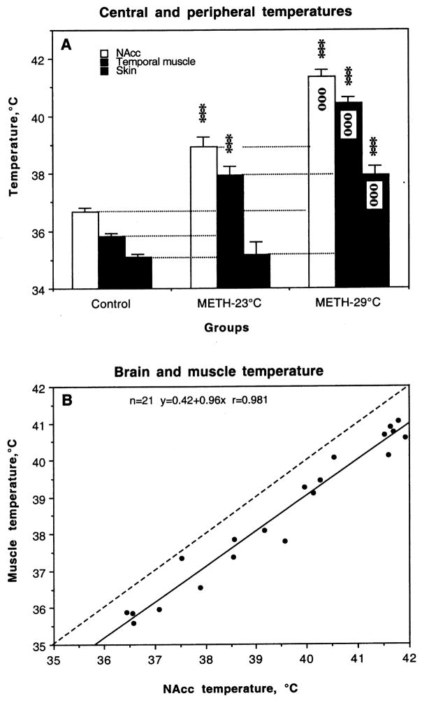
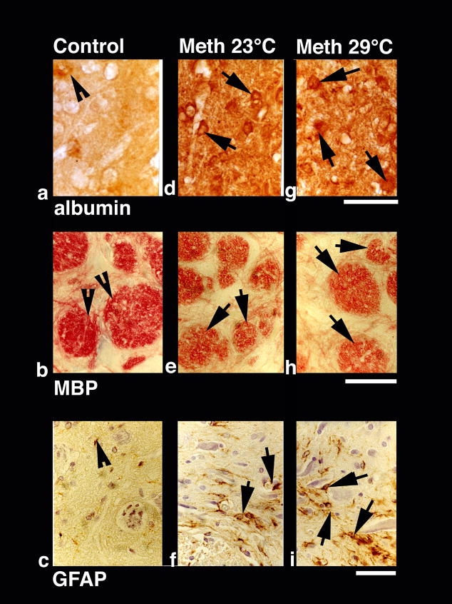
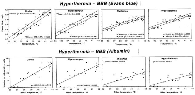
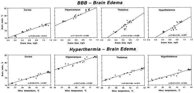
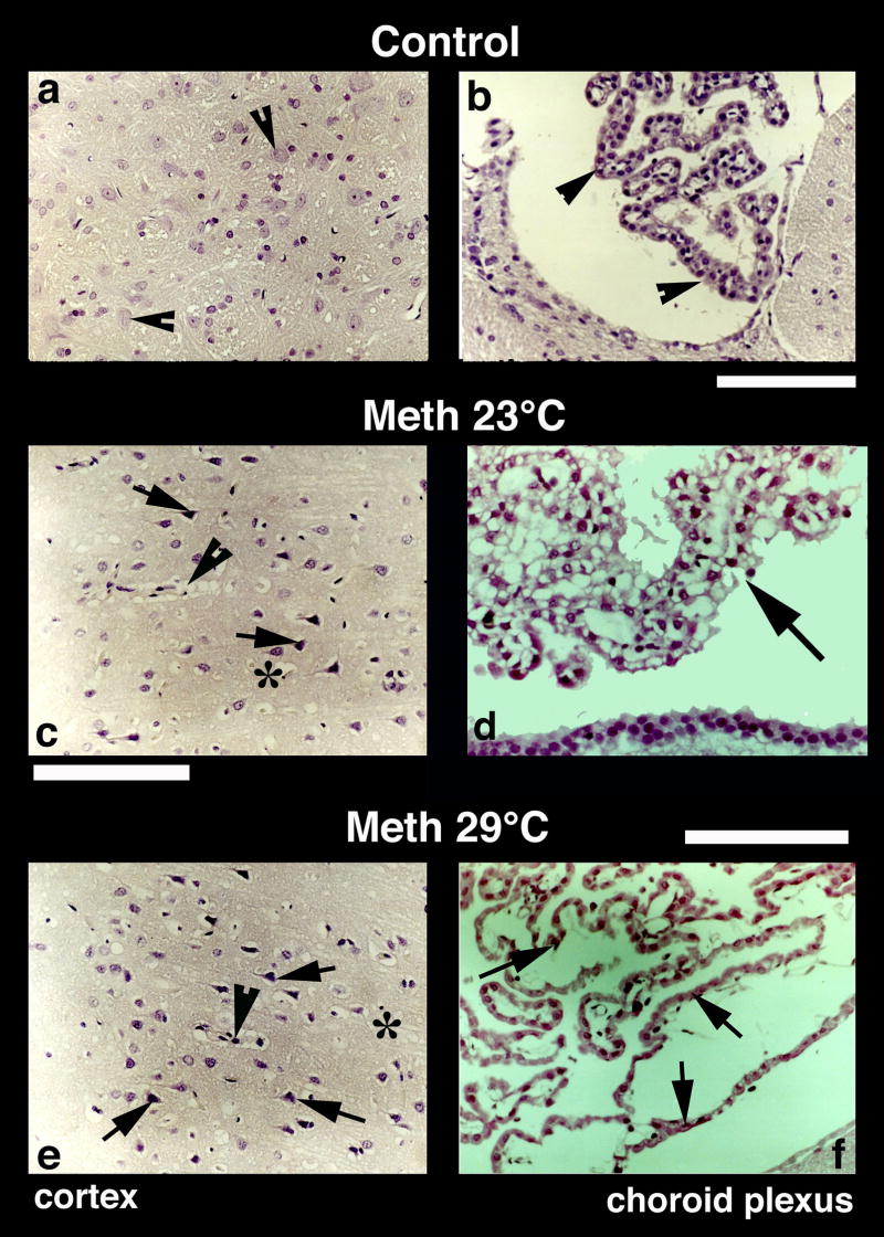
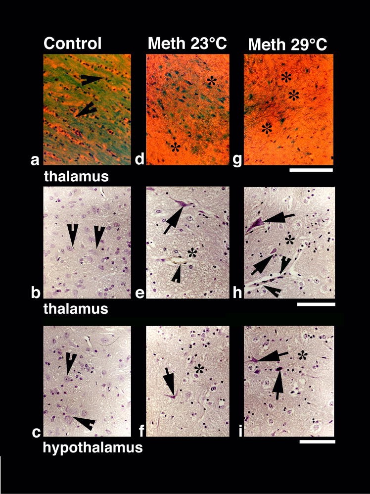
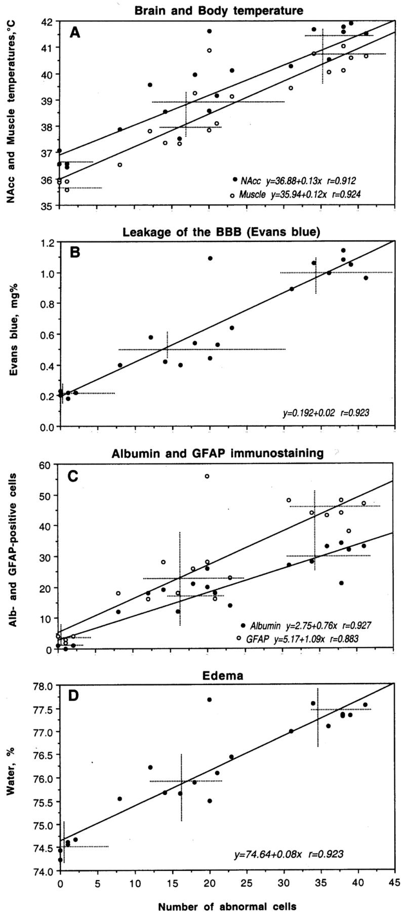
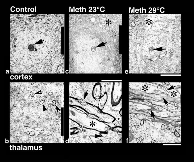
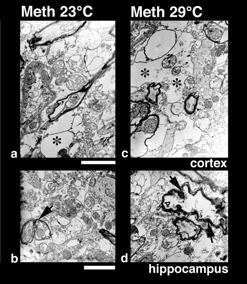
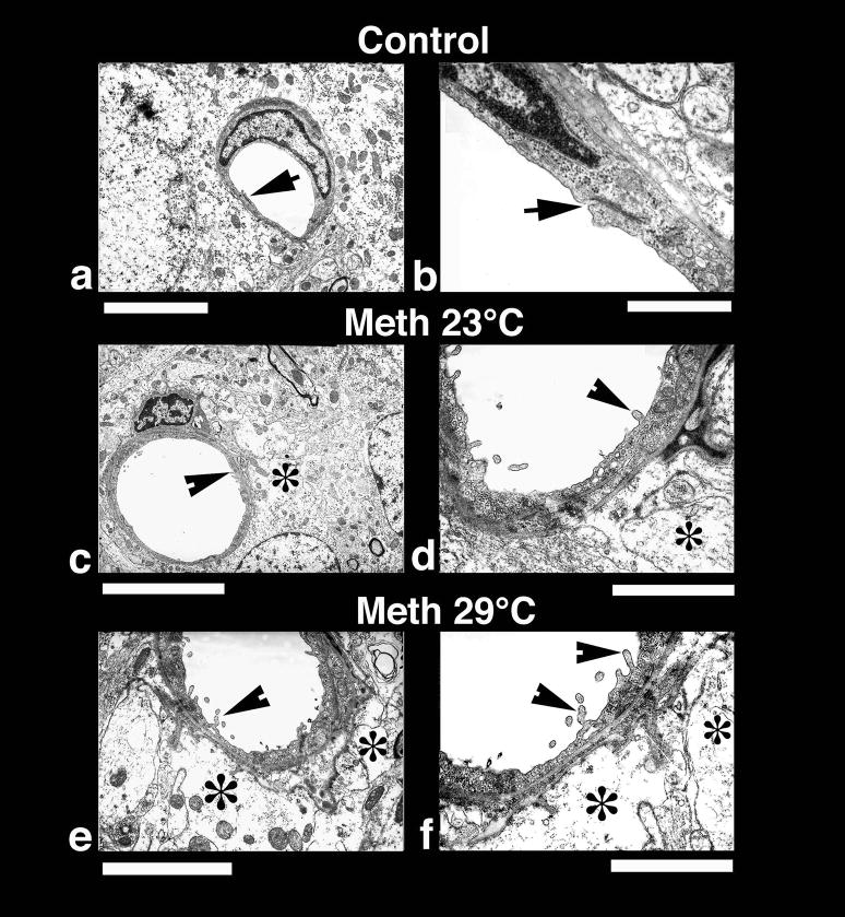
This study describes morphological abnormalities of brain cells during acute methamphetamine (METH) intoxication in the rat and demonstrates the role of hyperthermia, disruption of the blood-brain barrier (BBB) and edema in their development. Rats with chronically implanted brain, muscle and skin temperature probes and an intravenous (i.v.) catheter were exposed to METH (9 mg/kg) at standard (23 degrees C) and warm (29 degrees C) ambient temperatures, allowing for the observation of hyperthermia ranging from mild to pathological (38-42 degrees C). When brain temperature peaked or reached a level suggestive of possible lethality (>41.5 degrees C), rats were injected with Evans blue (EB), rapidly anesthetized, perfused, and their brains were taken for further analyses. Four brain areas (cortex, hippocampus, thalamus and hypothalamus) were analyzed for EB extravasation, water and electrolyte (Na(+), K(+), Cl(-)) contents, immunostained for albumin and glial fibrillary acidic protein (GFAP), and examined for neuronal, glial and axonal alterations using standard light and electron microscopy. These examinations revealed profound abnormalities in neuronal, glial, and endothelial cells, which were stronger with METH administered at 29 degrees C than 23 degrees C and tightly correlated with brain and body hyperthermia. These changes had some structural specificity, but in each structure they tightly correlated with increases in EB levels, the numbers of albumin-positive cells, and water and ion contents, suggesting leakage of the BBB, acutely developing brain edema, and serious shifts in brain ion homeostasis as leading factors underlying brain abnormalities. While most of these acute structural and functional abnormalities appear to be reversible, they could trigger subsequent cellular alterations in the brain and accelerate neurodegeneration-the most dangerous complication of chronic amphetamine-like drug abuse.


Figures

Fig. 1
Fig. 1
A. Changes in NAcc, muscle and skin temperatures induced by methamphetamine (METH, 9 mg/kg, sc) administered at standard (23°C) and warm (29°C) ambient temperatures. Asterisks show values significantly different from control (120 min after a sc saline injection at 23°C) and small circles show differences between two METH groups (***, ooo, p<0.001; Student’s t-test). B. Relationships between temperatures in the NAcc and temporal muscle assessed in all animals used in this study.
Fig. 2
Fig. 2
Immunohistochemical changes in albumin (a, d, g), myelin basic protein (MBP; b, e, g) and glial fibrillary acidic protein (GFAP; c, f, i) in control (left vertical panel) and METH-treated rats (23°: middle vertical panel and 29°C: right vertical panel). Compared to weak albumin immunoreactivity in control (a, arrowhead), METH-treated rats had stronger immonoreactivity (arrows in d and g). Expansion of neuropil and sponginess is also evident in the surrounding background. Bar (a,d,g) = 40 μm. In contrast to intense red myelin bundles and dense red fibers in control (b, arrowheads, b), diminution of red staining in the bundles and fibers (e, arrows) was seen in METH-treated rats (e). This degradation of MBP was most prominent in the rats treated with METH at 29° C (arrows, h). Bar (b, e, h) = 30 μm. GFAP immunostaining was prominent in METH-treated rats at 29° C (i, arrows) compared to 23°C (f, arrows). Control rats (c) occasionally show few GFAP positive astrocytes (arrowhead). Reactive astrocytes were located around the nerve cells and microvessels and were distributed in wide regions in the neuropil. Damaged neurons can also be seen in the background. Bar (c, f, i) = 40 μm.
Fig. 3
Fig. 3
Relationships between brain temperature (°C) and two parameters of BBB permeability (local concentrations of Evans blue, mg% and the number of Albumin-positive cells per section) in different brain structures during acute METH intoxication. Upper row also shows muscle temperature. Each graph shows a regression line, regression equation, and coefficient of correlation.
Fig. 4
Fig. 4
Relationships between permeability of the BBB (Evans blue, mg%) and brain edema (water,%) and between brain hyperthermia (NAcc temperature, °C) and edema in different brain structures during acute METH intoxication. Each graph shows a regression line, regression equation, and coefficient of correlation.
Fig. 5
Fig. 5
Nissl-stained sections from the cerebral cortex (left panel) and choroid plexus (right panel) from control (a, b), METH-23°C (c, d) and METH-29°C (e, f) treated rats. Most of the nerve cells in the control cortex are healthy with a distinct nucleus in the center. Only a few nerve cells show a condensed cytoplasm (arrowheads, a). On the other hand, dark and distorted neurons were frequent in METH-treated rats (c and e; arrows). Sponginess (*) and perivascular edema (arrowheads; c, e) are frequent in METH-treated rats; the changes are more pronounced at 29° C (e) compared to 23° C (c). Bars (a, c, e) = 50 μm. Nissl-stained choroid epithelial cells in control (b) show compact and densely packed epithelial cells with a distinct cell nucleus (arrow, b). METH treatment at 23° C resulted in mild degeneration of choroid epithelium (arrows, d). The epithelial cell nucleus also appears to be disintegrated (d). These degenerating changes in the choroidal epithelium is most pronounced in the rat that received METH at 29° C (arrows, f). Bars (b = 50 μm; d = 40 μm, f = 60 μm).
Fig. 6
Fig. 6
Luxol Fast blue (upper panel) and Nissl staining (middle and lower panels) in control (a–c), METH-23°C (d–f) and METH 29°C (g–i) treated rats. Dense myelinated axons and fibers (arrow heads, a) stained by Luxol Fast Blue seen in control (a) diminished in the METH-23°C group (d) and generally lost in METH-29°C group (g). Sponginess and edema (*) is seen in the background of Luxol Fast Blue-stained elements (d, g). Bar = 50 μm. Nissl-stained nerve cells show pronounced degenerative changes in the thalamus (middle panel) and hypothalamus (lower panel) that are stronger in METH-29°C (arrows, h, i) than METH=23°C group (arrows, e, f). Control animals showed healthy neurons with a distinct nucleus in both the thalamus (arrow heads b) and hypothalamus (arrow heads c). Perivascular edema (arrowhead e) and sponginess (*) are most marked in the METH-29°C group (h, i). Bars (b, e, h = 40 μm; c, f, i = 40 μm).
Fig. 7
Fig. 7
The relationships between cortical neural damage and several other functional parameters during acute METH intoxication. Abscissas in each graph shows the number of damaged cells encountered in cortex and ordinates show, respectively, NAcc and muscle temperatures (A), concentrations of Evans blue (B), the numbers of Albumin- and GFAP-positive neurons (C) and tissue water content (D). Each graph shows a regression line (s), regression equation (s), and coefficient(s) of correlation (r). Hatched lines show mean values.
Fig. 8
Fig. 8
Low-power transmission electron micrograph from the cortex (upper panel) and thalamus (lower panel) showing neuronal nuclear (a, c, e) and axonal changes (b, d, f) in control (a, b), METH-23° C (c, d) and METH-29° C (e, f) groups. The neuronal nucleus in control rat shows a smooth nuclear envelope with a dark granular karyoplasm containing a central nucleolus (a, arrow). The nerve cell cytoplasm is compact and condensed without any vacuoles. On the other hand, a less electron-dense karyoplasm with an eccentric nucleolus showing degenerative changes is seen in the METH-23°C group (c, arrow). The nuclear membrane showed irregular foldings and vacuolation (*) in the neuropil including cytoplasm. These changes in the cell nucleus were much more aggravated in rats treated with METH at 29°C (e); degeneration of the nuclear membrane and surrounding neurnal cytoplasm is clearly evident in this slice. The nucleolus was further degenerated (arrow) and became more eccentric (e). Bar: a–c = 1 μm. Axonal changes in the thalamus of METH-treated rats at 23°C (d) show profound myelin vesiculation (arrow) and edematous swelling (*, d). These changes were stronger in rats treated with METH at 29° C (f). In this group, the myelin vesiculation (arrows) and degeneration of axons were clearly evident (*, f). On the other hand, normal rats exhibited a compact neuropil with normal myelinated axons (arrow heads, b). Signs of vacuolation and edema are largely absent in control group (a, b). Bars: b = 1500 nm, d = 800 nm; f = 600 nm.
Fig. 9
Fig. 9
High-power transmission electron micrograph of neuropil from the cortex (upper panel) and hippocampus (lower panel) in rats treated with METH at 23° C (a, b) and 29° C (c, d). Vacuolation, edema (*) and degeneration of myelin are much more extensive and widespread in both the cortex (c) and hippocampus (d) in rats treated with METH at 29°C compared to 23°C (a, b). However, mitochondria in the neuropil are largely preserved showing normal cristae indicating that these membrane disruption and vacuolation are not due to fixation artifacts (for details see text). Bar: a, c = 600 nm; c, d = 800 nm.
Fig. 10
Fig. 10
Low-power (left panel) and High-power (right panel) transmission electronmicrographs (left panel) showing a cerebral capillary and the surrounding neuropil in control (a and b) and METH-treated brain (c and d, 23°; e and f 29° C). A normal cerebral capillary has a smooth luminal surface and a compact, dense neuropil surrounding it and normal tight junctions (a, arrow). The normal capillary also has distinct tight junctions (b, arrow) and the underlying glial cells (astrocyte) do not exhibit any apparent signs of perivascular edema (b). METH treatment at 23°C resulted in endothelial cell reaction and swelling of the perivascular astrocyte (*, c). The endothelial luminal surface exhibited few distinct bleb formations (arrow head) indicating the process of enhanced vesicular transport or alterations in membrane transport properties (d, for details see text). Swollen perivascular astrocytes and its processes (*) are evident in this METH-treated rat (d). These ultratstructural changes, e.g., bleb formation and perivascular edema, were much more aggravated in the rat after METH treatment at 29°C (e, f). Thus, spreading out of small membrane vesicles and elongated bleb formation could be seen in this group (arrow heads). Swelling of astrocytes (*) and disintegration of astrocytic cytoplasm indicating water filled cells are clearly visible (*, f). The endothelial cell cytoplasm in METH-treated rats is much more condensed (d, f) compared to control (a, b). Bars: a, c = 1 μm, e = 2 μm,; b = 500 nm; d = 800 nm; f = 600 nm.

References

    1. Alberts DS, Sonsalla PK. Methamphetamine-induced hyperthermia and dopaminergic neurotoxicity in mice: Pharmacological profile of protective and nonprotective agents. J Pharmacol Exp Ther. 1995;275:1104–1114. - PubMed
    1. Ali SF, Newport GD, Slikker W. Methamphetamine-induced dopaminergic toxicity in mice: Role of environmental temperature and pharmacological agents. Ann NY Acad Sci. 1996;801:187–198. - PubMed
    1. Bechtold DA, Brown IR. Induction of Hsp27 and Hsp32 stress proteins and vimentin in glial cells of the rat hippocampus following hyperthermia. Neurochem Res. 2003;28:1163–1173. - PubMed
    1. Bekay L, Lee JC, Lee GC, Peng GR. Experimental cerebral concussion: An electron microscopic study. J Neurosurg. 1977;47:525–531. - PubMed
    1. Blezer E. Techniques for measuring the blood-brain barrier integrity. In: de Vries E, Prat A, editors. the Blood-Brain Barrier and Its Microenvironment. New York: Taylor & Francis; 2005. pp. 441–456.

Publication types

MeSH terms