Review
. 2008 Oct;31(10):529-37.
doi: 10.1016/j.tins.2008.07.003. Epub 2008 Sep 4.

The healthy cell bias of estrogen action: mitochondrial bioenergetics and neurological implications

Affiliations
Review

The healthy cell bias of estrogen action: mitochondrial bioenergetics and neurological implications

Roberta Diaz Brinton. Trends Neurosci. 2008 Oct.

Abstract

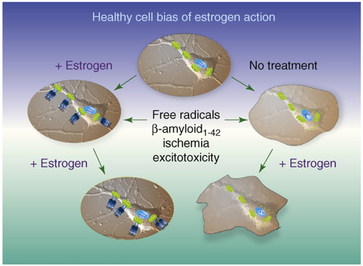
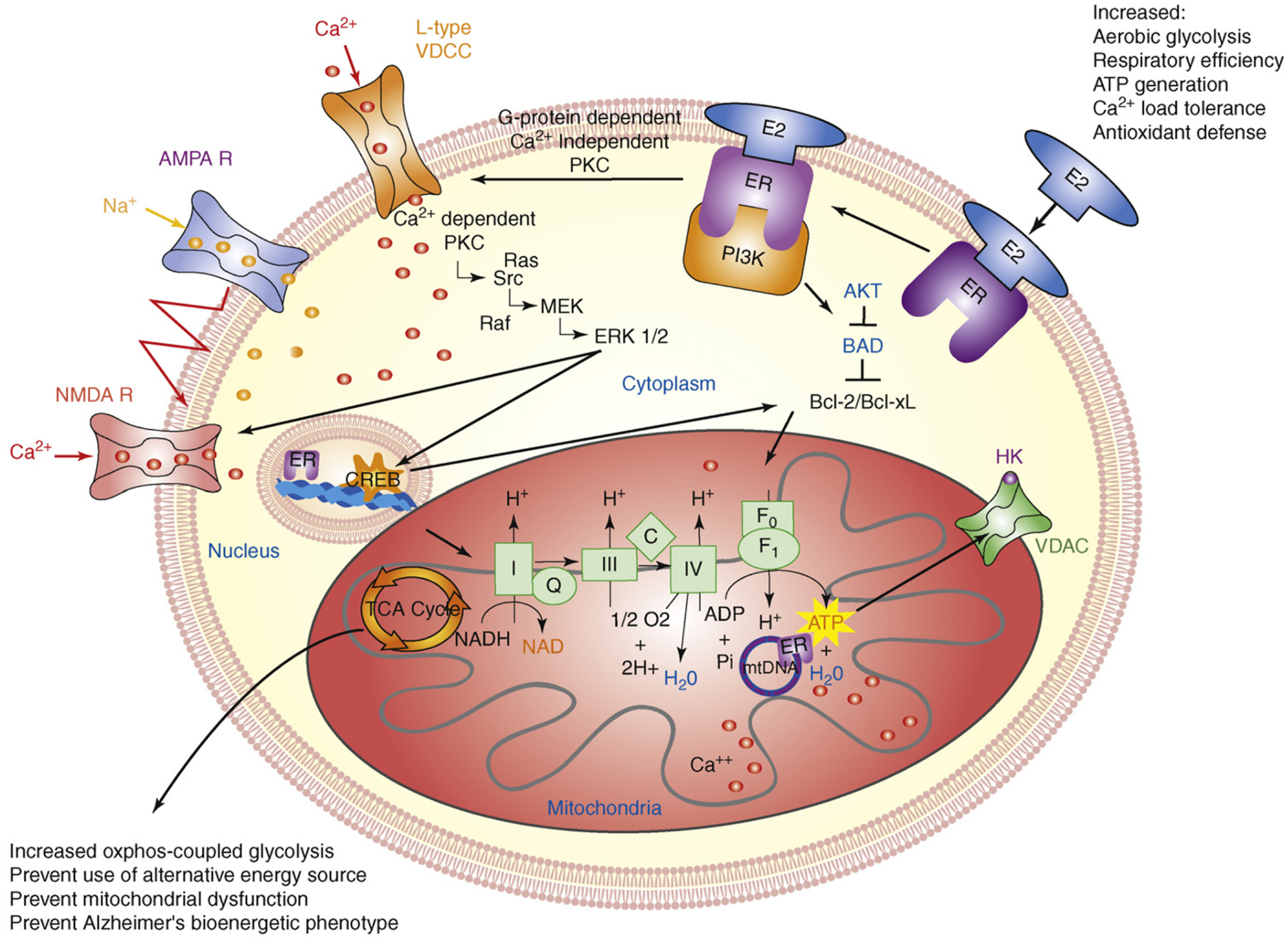
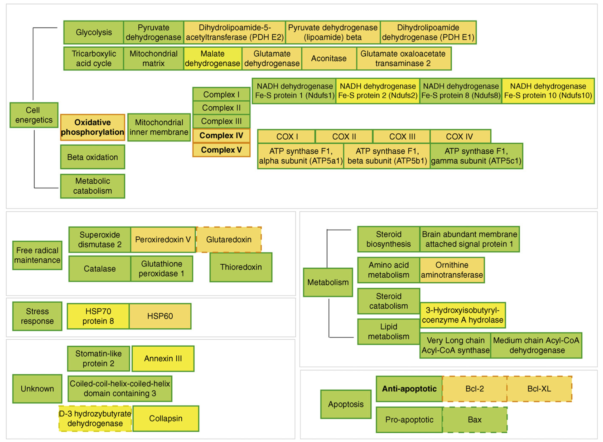
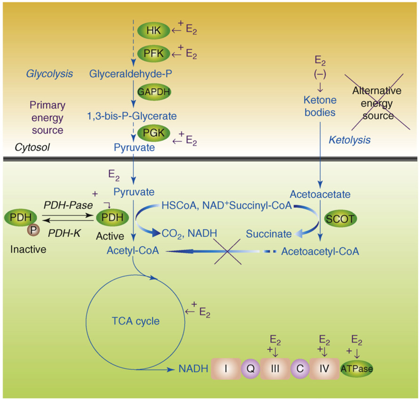
The 'healthy cell bias of estrogen action' hypothesis examines the role that regulating mitochondrial function and bioenergetics play in promoting neural health and the mechanistic crossroads that lead to divergent outcomes following estrogen exposure. Estrogen-induced signaling pathways in hippocampal and cortical neurons converge upon the mitochondria to enhance aerobic glycolysis coupled to the citric acid cycle, mitochondrial respiration and ATP generation. Convergence of estrogen-induced signaling onto mitochondria is also a point of vulnerability when activated in diseased neurons which exacerbates degeneration through increased load on dysregulated calcium homeostasis. As the continuum of neurological health progresses from healthy to unhealthy so too do the benefits of estrogen or hormone therapy. The healthy cell bias of estrogen action hypothesis provides a lens through which to assess disparities in outcomes across basic and clinical science and on which to predict outcomes of estrogen interventions for sustaining neurological health and preventing age-associated neurodegenerative diseases such as Alzheimer's.


Figures

Figure 1.
Figure 1.
Healthy cell bias of estrogen action evidence from basic to clinical science indicates that neurons and women treated with estrogen before exposure to neurodegenerative insult prevents neural demise. In stark contrast, basic and clinical evidence further indicates that exposure to estrogen following neurodegenerative insult can result in an exacerbation of neurological demise. Estrogen regulation of calcium signaling and mitochondrial function play key roles in determining the outcome of estrogen exposure. Figure modified from Ref. [1].
Figure 2.
Figure 2.
Estrogen mechanisms of action converge upon the mitochondria. Estrogen (17β-estradiol; E2) binding to a membrane-associated estrogen receptor (ER) undergoes a protein-protein interaction with the regulatory subunit of PI3K, p85, to activate the divergent but coordinated activation of the Akt and MAPk signaling cascades. These E2-induced signaling pathways in hippocampal and cortical neurons converge upon the mitochondria to enhance glucose uptake and metabolism, aerobic glycolysis and pyruvate dehydrogenase to couple aerobic glycolysis to acetyl-CoA production and tricarboxylic acid cycle (TCA) -coupled oxidative phosphorylation and ATP generation. In parallel, E2 increases antioxidant defense and antiapoptotic mechanisms. Estrogen receptors at the membrane, in mitochondria and within the nucleus are well positioned to regulate coordinated mitochondrial and nuclear gene expression required for optimal bioenergetics. Enhancing and sustaining glycolysis, aerobic metabolism and mitochondrial function would be predicted to prevent the shift to alternative fuel sources and the hypometabolism characteristic of Alzheimer’s disease. Figure modified from Ref. [2].
Figure 3.
Figure 3.
Overview of 17β-estradiol (E2) regulation of female rat brain mitoproteome in vivo. Results of the functional proteomic analysis of the brain mitoproteome were combined with a bioinformatic assessment of the brain mitoproteome regulated by E2. Proteins with known responses to E2 were separated into functional subgroups based on common mitochondrial ontology. Orange represents upregulation and yellow represents downregulation. Filled boxes are based on results of Nilsen and Irwin and colleagues [25]. Dashed boxes are derived from published literature (reviewed in Ref. [25]). Bold lettering represents altered activity. E2 significantly increased key components of the cellular energetic machinery including proteins involved in the tricarboxylic acid cycle and oxidative phosphorylation. Further, E2 increased expression of antioxidant enzymes and antiapoptotic proteins. Collectively, the data indicate a comprehensive regulation of mitochondrial function by E2 which increases key elements in the tricarboxylic acid cycle, pyruvate metabolism, mitochondrial oxidative phosphorylation, respiratory efficiency and ATP generation while reducing free radical leak and oxidative damage.
Figure 4.
Figure 4.
Estrogen (E2) promotes glycolysis and glycolytic-coupled tricarboxylic acid cycle (TCA) function, mitochondrial respiration and ATP generation to prevent a switch to ketone bodies as an alternative fuel source. E2 increases key enzymes in the glycolytic pathway to promote generation of pyruvate and its conversion by pyruvate dehydrogenase (PDH) to acetyl-CoA to initiate and sustain the TCA cycle. Under metabolically challenging conditions (i.e. starvation, aging and neurodegeneration), neurons can utilize acetyl-CoA generated from ketone body metabolism (ketolysis), produced by the liver or under conditions of starvation in neighboring glial cells. This latter pathway is much less efficient and can inhibit residual glycolysis. In AD, there is a generalized shift toward use of an alternative fuel, ketone bodies, and away from glycolytic energy production. Estrogen enhances glucose uptake into the brain glycolytic/pyruvate/acetyl-CoA pathway to generate electrons required for oxidative phosphorylation and ATP generation. Collectively, estrogen enhancement of glucose metabolism and aerobic glycolysis promotes and sustains utilization of glucose as the primary fuel source of the brain, thereby preventing the shift to alternative fuels such as ketone bodies which is characteristic of Alzheimer’s disease.

References

    1. Brinton RD (2005) Investigative models for determining hormone therapy-induced outcomes in brain: evidence in support of a healthy cell bias of estrogen action. Ann. N. Y. Acad. Sci 1052, 57–74 - PubMed
    1. Morrison JH et al. (2006) Estrogen, menopause, and the aging brain: how basic neuroscience can inform hormone therapy in women. J. Neurosci 26, 10332–10348 - PMC - PubMed
    1. Wise PM (2006) Estrogen therapy: does it help or hurt the adult and aging brain? Insights derived from animal models. Neuroscience 138, 831–835 - PubMed
    1. Singh M et al. (2008) Estrogens and progesterone as neuroprotectants: what animal models teach us. Front. Biosci 13, 1083–1089 - PMC - PubMed
    1. Nilsen J and Brinton RD (2002) Impact of progestins on estrogen-induced neuroprotection: synergy by progesterone and 19-norprogesterone and antagonism by medroxyprogesterone acetate. Endocrinology 143, 205–212 - PubMed

Publication types