Review
. 2008 Oct;18(10):474-85.
doi: 10.1016/j.tcb.2008.08.002. Epub 2008 Sep 4.

Phagosome maturation during the removal of apoptotic cells: receptors lead the way

Affiliations
Review

Phagosome maturation during the removal of apoptotic cells: receptors lead the way

Zheng Zhou et al. Trends Cell Biol. 2008 Oct.

Abstract

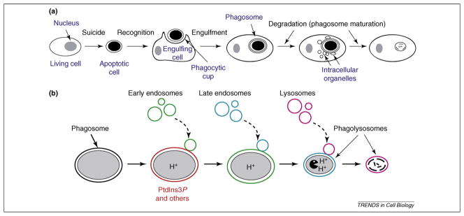
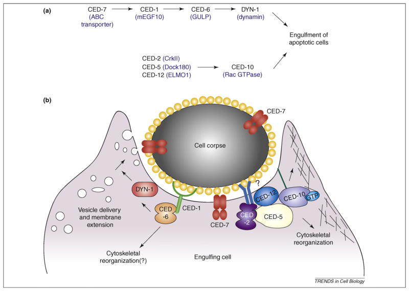
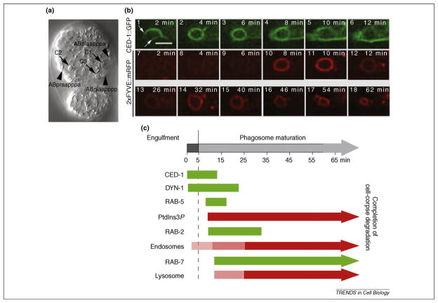
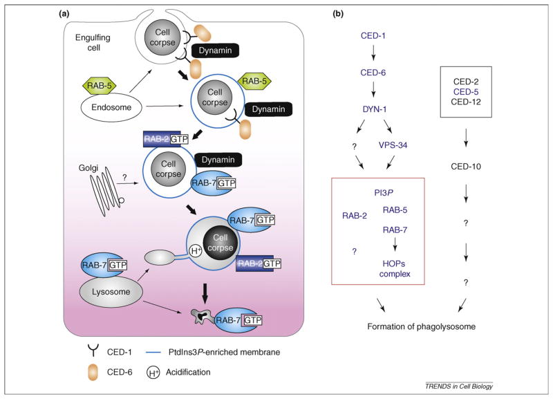
In metazoan organisms, cells undergoing apoptosis are rapidly engulfed and degraded by phagocytes. Defects in apoptotic-cell clearance result in inflammatory and autoimmune responses. However, little is known about how apoptotic-cell degradation is initiated and regulated and how different phagocytic targets induce different immune responses from their phagocytes. Recent studies in mammalian systems and invertebrate model organisms have led to major progress in identifying new factors involved in the maturation of phagosomes containing apoptotic cells. These studies have delineated signaling pathways that promote the sequential incorporation of intracellular organelles to phagosomes and have also discovered that phagocytic receptors produce the signals that initiate phagosome maturation. Here, we discuss these exciting new findings, focusing on the mechanisms that regulate the interactions between intracellular organelles and phagosomes.


Figures

Figure 1
Figure 1
Apoptotic cells are engulfed by phagocytes and, subsequently, digested inside phagosomes in metazoans. (a) The process of apoptotic-cell engulfment and degradation. Cell death proceeds in four stages: (i) the specification of a death fate (not shown here); (ii) cell-death execution; (iii) recognition and engulfment by phagocytes; and (iv) degradation within phagosomes. During phagosome maturation, a variety of intracellular organelles are sequentially incorporated into phagosomes. (b) The sequential incorporation of early endosomes, late endosomes and lysosomes to phagosomes drives phagosome maturation. These vesicles (identities indicated by particular colors) are recruited to the phagosomal surfaces and then fuse with phagosomes, providing the phagosome with a variety of protein and lipid materials. Phagosome luminal PH starts to decrease after the completion of engulfment and reaches the lowest level when a phagosome evolves into a phagolysosome. A phagolysosome gradually decreases in size and eventually disappears.
Figure 2
Figure 2
The C. elegans engulfment genes and their molecular functions. (a) Two parallel and partially redundant pathways that control the engulfment of apoptotic cells in C. elegans. Members of each pathway were identified from genetic screens for mutations that impair cell-corpse removal. Each gene was placed in one of the two pathways based on the results of epistasis grouping assays and phenotype characterizations. The order of genes in each pathway was determined by epistasis analyses (for details, see text and Ref. [54]). The identities of the corresponding mammalian homologs are indicated inside parentheses. (b) The molecular functions and protein–protein interactions of the eight genes listed in (a). Yellow spheres coating the cell corpse represent the ‘eat me’ signal presented by cell corpses to attract engulfing cells. Among the eight proteins, CED-7 is the only one that functions in both dying and engulfing cells. All other proteins function specifically in engulfing cells for cell-corpse engulfment. CED-2, CED-5 and CED-12 form a protein complex under the plasma membrane to regulate the CED-10 Rac GTPase, which promotes cytoskeletal reorganization and pseudopod extension during engulfment. CED-5 and CED-12 function as a bipartite guanine-nucleotide-exchange factor that activates CED-10. CED-10 associates with the plasma membrane through its prenylated C terminus. Two deep blue lines, which represent a transmembrane receptor that interacts with an ‘eat me’ signal molecule and a nearby question mark indicate that the transmembrane receptor(s) expected to interact with the SH2 domain of CED-2 with its intracellular domain and activate the CED-2, -5 and –12 complex has not been identified. In the other pathway, CED-7 mediates the presentation of an ‘eat me’ signal on the surface of apoptotic cells, and might possess a different engulfment-promoting activity on the surface of engulfing cells. CED-1 is a phagocytic receptor that uses its extracellular domain to recognize the ‘eat me’ signal, clusters around the cell corpse, and uses its intracellular domain for downstream signaling. CED-6 interacts with CED-1 and functions as the adaptor of CED-1. DYN-1 is localized to the surface of pseudopods and phagosomes in a manner dependent on the functions of CED-1, -6 and -7. DYN-1 mediates the signaling of CED-1 to promote the delivery and incorporation of intracellular vesicles to pseudopods through ‘focal phagocytosis’, a mechanism that provides membrane material to support pseudopod extension. DYN-1 also mediates CED-1 signaling to promote phagosome maturation. An additional activity of CED-1 and CED-6 in actin reorganization, possibly mediated by CED-10, has also been suggested. For review, see main text and Refs [9,50,54,62,68,69]. Figure adapted, with permission, from Ref. [61].
Figure 3
Figure 3
Time-lapse recording of the engulfment and degradation of apoptotic cells and the dynamic phagosome localization pattern of multiple essential factors in C. elegans embryos. (a) DIC image of an embryo at 3330 min post the first embryonic division. Anterior is to the top. Ventral faces readers. Arrowheads indicate the three ventral hypodermal cells that engulf apoptotic cells C1, C2 and C3 (arrows); the identity of each engulfing cell is labeled. (b) Real-time observation of the engulfment and degradation of cell-corpse C3. Time-lapse images of the co-expressed CED-1–GFP (1–7) and 2xFYVE–mRFP reporters (8–18) around C3 in a wild-type embryo. CED-1–GFP labels pseudopods, enabling the visualization of the pesudopod extension and fusion along C3. 2xFYVE–mRFP, which associates with PtdIns3P, labels phagosomes throughout their entire duration, and is used here to demonstrate the formation and the gradual shrinkage of a phagosome containing C3, a process that represents phagosome maturation and the degradation of cargo. 0 min: the time point that the engulfing cell starts to extend pseudopods around C3. White arrows indicate the edge of pseudopods. Scale bar: 3 μm. (c) A summary of the temporal order of the phagosome localization of multiple essential factors for phagosome maturation and of the incorporation of endosomes and lysosomes, using GFP or mRFP-fusion reporters. Data represent mean values obtained from time-lapse recording monitoring the engulfment and degradation of multiple C1, C2, and C3 cell corpses. ‘0 min’ represents the time point when budding pseudopods are first detected by CED-1–GFP. The pale pink colors used for endosomal and lysosomal incorporation into phagosomes indicate that a very weak signal was detected at an early stage and signal intensities gradually increase. For review see Refs [62,69,73].
Figure 4
Figure 4
Intracellular-vesicle-transport factors that are involved in phagosome maturation and the two genetic pathways that control degradation of apoptotic cells in C. elegans (a) In C. elegans, phagocytic receptor CED-1, large GTPase Dynamin and small GTPases, Rab5, Rab2 and Rab7, sequentially localize to the phagocytic cup, nascent phagosomes and phagolysosomes to mediate phagosome maturation through different stages. At each stage, endosomes and lysosomes fuse with maturing phagosomes. Golgi apparatus might also take part in the regulation of phagosome maturation through the activity of Rab2. (b) The two parallel genetic pathways that were previously identified to regulate cell-corpse engulfment in C. elegans are now found to also control phagosome maturation. Proteins, the names of which are in blue, are those that have been found to be required for degradation of apoptotic cells using loss-of-function mutations. Proteins in black are in the engulfment pathways that have not been tested for their phagosome-maturation activity. The positions in the pathway of those protein and lipid molecules listed in the red box have not been fully determined (for details, see main text and Table 1). The two pathways would eventually converge on the formation of a phagolysosome, after which step the phagosome degrades its content irreversibly.

Similar articles

Cited by

References

    1. Caron E, Hall A. Phagocytosis. In: Marsh M, editor. Endocytosis. Oxford University Press; 2001. pp. 58–77.
    1. Vieira OV, et al. Phagosome maturation: aging gracefully. Biochem J. 2002;366:689–704. - PMC - PubMed
    1. Erwig LP, Henson PM. Immunological consequences of apoptotic cell phagocytosis. Am J Pathol. 2007;171:2–8. - PMC - PubMed
    1. Vaux DL, Korsmeyer SJ. Cell death in development. Cell. 1999;96:245–254. - PubMed
    1. Henson PM, Hume DA. Apoptotic cell removal in development and tissue homeostasis. Trends Immunol. 2006;27:244–250. - PubMed

Publication types

MeSH terms

Substances

LinkOut - more resources