Review
. 2008 Oct;18(10):467-73.
doi: 10.1016/j.tcb.2008.08.001. Epub 2008 Sep 4.

Apoptosis-induced compensatory proliferation. The Cell is dead. Long live the Cell!

Affiliations
Review

Apoptosis-induced compensatory proliferation. The Cell is dead. Long live the Cell!

Yun Fan et al. Trends Cell Biol. 2008 Oct.

Abstract

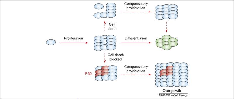
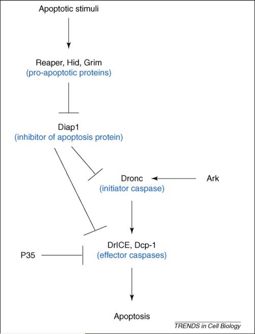
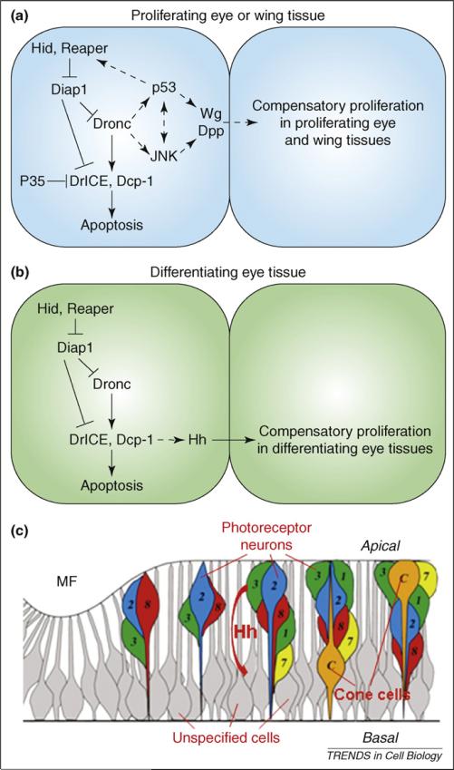
In multi-cellular organisms, activation of apoptosis can trigger compensatory proliferation in surrounding cells to maintain tissue homeostasis. Genetic studies in Drosophila have indicated that distinct mechanisms of compensatory proliferation are employed in apoptotic tissues of different developmental states. In proliferating eye and wing tissues, the initiator caspase Dronc coordinates cell death and compensatory proliferation through the Jun N-terminal kinase and p53. The mitogens Decapentaplegic and Wingless are induced in this process. By contrast, in differentiating eye tissues, the effector caspases DrICE and Dcp-1 activate the Hedgehog signaling pathway to induce compensatory proliferation. In this review, we summarize these findings and discuss how activation of apoptosis is linked to the process of compensatory proliferation. The developmental and pathological relevance of compensatory proliferation is also discussed.


Figures

Figure 1
Figure 1
Model of compensatory proliferation in response to apoptosis. In multi-cellular organisms, morphogenesis is composed of patterned cell proliferation followed by cell differentiation (green). Compensatory proliferation can be induced in response to cell loss to maintain tissue homeostasis. If execution of cell death is blocked, for example by expression of P35, an effector-caspase inhibitor in Drosophila, ‘undead cells’ (red) continue to secrete signals to induce proliferation, resulting in tissue overgrowth.
Figure 2
Figure 2
The apoptotic pathway in Drosophila. In response to apoptotic stimuli, expression of the pro-apoptotic genes hid, reaper and grim releases the initiator caspase Dronc from inhibition by Diap1. Free Dronc then associates with the adaptor protein Ark and proteolytically processes the effector caspases DrICE and Dcp-1 to induce apoptosis.
Figure 3
Figure 3
Two modes of apoptosis-induced compensatory proliferation in proliferating and differentiating eye tissues in Drosophila. (a) Schematic outline of the late-third instar eye imaginal disc. Anterior is to the left. The developing larval eye disc is composed of the anterior proliferating tissue (red) and the posterior differentiating tissue (green), which are separated by G1-arrested cells, the morphogenetic furrow (MF) and the second mitotic wave (SMW). The eyeless (ey) promoter is expressed in the anterior part (red); the GMR promoter is expressed in the posterior part (green) and in the SMW. (b) An eye imaginal disc from a 3rd instar wild-type larva labeled with BrdU as the proliferation marker. The anterior half of the disc [red in (a)] is heavily proliferating. Posterior to the SMW [green in (a)], proliferation has stopped and cells are differentiating into photoreceptor neurons and accessory cell types. The SMW is marked by a white arrowhead. (c) Co-expression of hid and P35 under ey control (ey-Gal4, UAS-hid, UAS-P35) triggers overgrowth of the anterior compartment [red in (a); compare with (b)]. The white bar indicates the extent of overgrowth [compare to (b)]. (d) Expression of hid under GMR control (GMR-hid) triggers compensatory proliferation (white arrow) posterior to the SMW [green in (a); compare with (b)]. Part (a) adapted, with permission, from Ref. [13].
Figure 4
Figure 4
Models of distinct mechanisms of compensatory proliferation in proliferating versus differentiating tissues in Drosophila. (a,b) Molecular mechanisms of apoptosis-induced compensatory proliferation in proliferating tissues (a) and differentiating tissues (b). An apoptotic cell (left) and a cell that is induced to undergo compensatory proliferation (right) are shown. Dashed arrows indicate unknown interactions. The scaffolding protein Ark is omitted for clarity (Figure 2). (c) Schematic outline of various cell types in the differentiating eye tissue. Hh produced in apical dying photoreceptor neurons (numbered colored cells) non-autonomously induces cell-cycle re-entry of basally-located unspecified cells (gray cells). ‘Apical’ and ‘basal’ refer to the apical and basal sides of the disc. The SMW is omitted for clarity. Part (c) adapted, with permission, from Ref. [13].

Similar articles

Cited by

References

    1. Danial NN, Korsmeyer SJ. Cell death: critical control points. Cell. 2004;116:205–219. - PubMed
    1. Haynie JL, Bryant PJ. The effects of X-rays on the proliferation dynamics of cells in the imaginal disc of Drosophila melanagaster. Rouxs Arch. Dev. Biol. 1977;183:85–100. - PubMed
    1. James AA, Bryant PJ. A quantitative study of cell death and mitotic inhibition in γ-irradiated imaginal wing discs of Drosophila melanogaster. Radiat. Res. 1981;87:552–564. - PubMed
    1. Milan M, et al. Developmental parameters of cell death in the wing disc of Drosophila. Proc. Natl. Acad. Sci. U. S. A. 1997;94:5691–5696. - PMC - PubMed
    1. Valentin-Vega YA, et al. The intestinal epithelium compensates for p53-mediated cell death and guarantees organismal survival. Cell Death Differ. 2008. DOI: 10.1038/cdd.2008.109 (http://www.nature.com/cdd) - PMC - PubMed

Publication types