. 2008 Oct 25;380(2):338-43.
doi: 10.1016/j.virol.2008.07.029. Epub 2008 Sep 6.

Organization of the mouse RNA-specific adenosine deaminase Adar1 gene 5'-region and demonstration of STAT1-independent, STAT2-dependent transcriptional activation by interferon

Affiliations

Organization of the mouse RNA-specific adenosine deaminase Adar1 gene 5'-region and demonstration of STAT1-independent, STAT2-dependent transcriptional activation by interferon

Cyril X George et al. Virology. .

Abstract

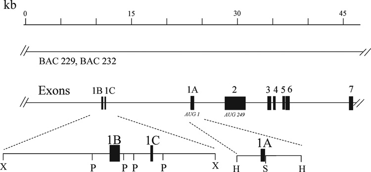
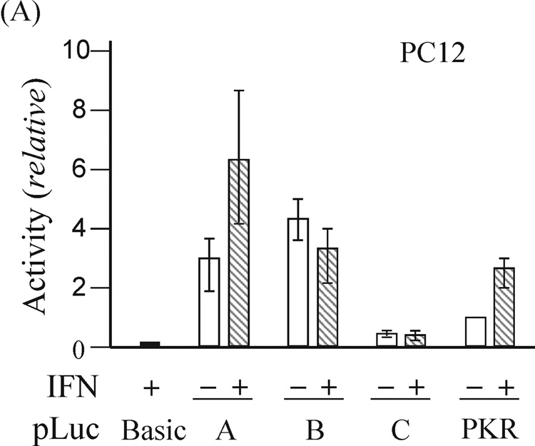
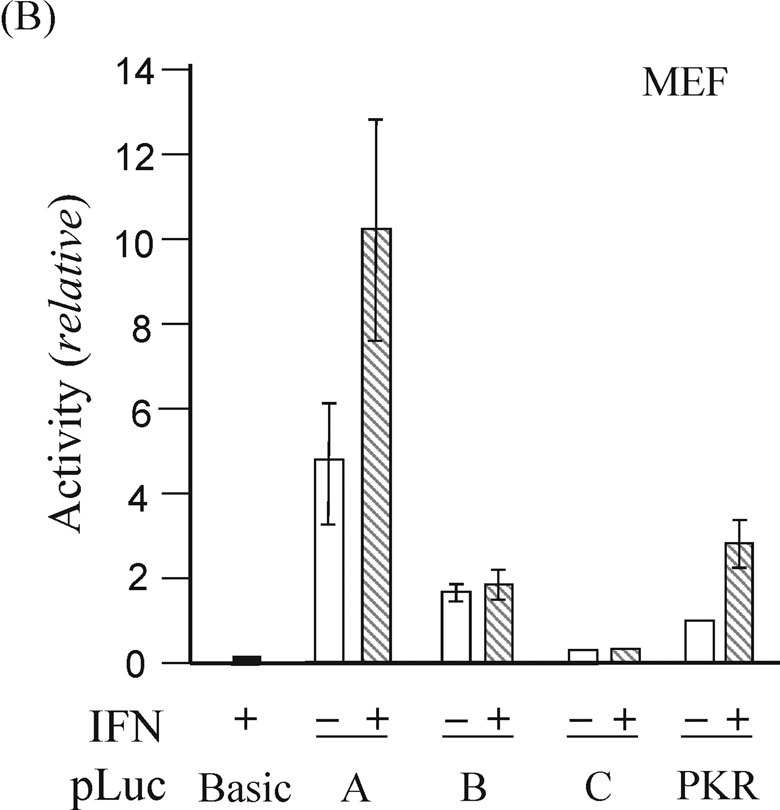
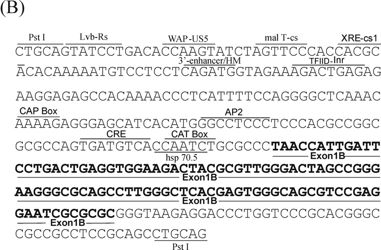
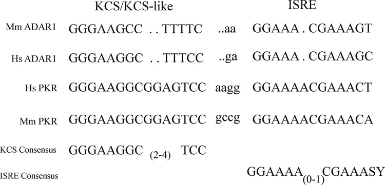
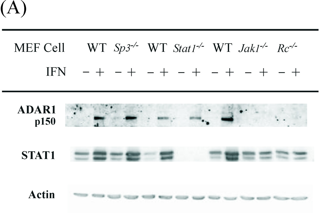
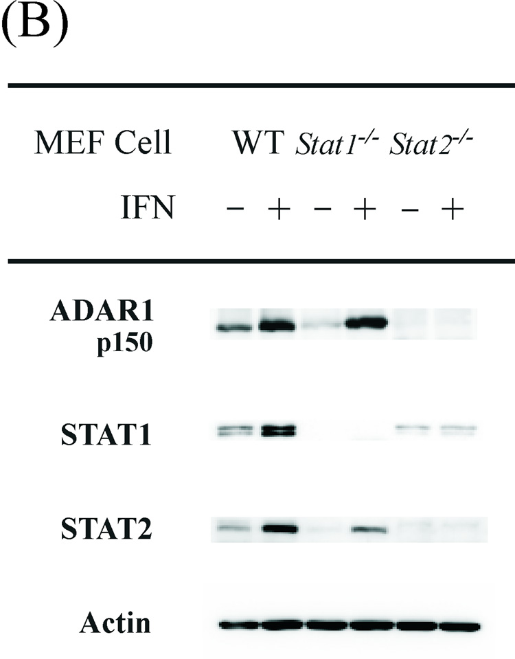
The p150 form of the RNA-specific adenosine deaminase ADAR1 is interferon-inducible and catalyzes A-to-I editing of viral and cellular RNAs. We have characterized mouse genomic clones containing the promoter regions required for Adar1 gene transcription and analyzed interferon induction of the p150 protein using mutant mouse cell lines. Transient transfection analyses using reporter constructs led to the identification of three promoters, one interferon-inducible (P(A)) and two constitutively active (P(B) and P(C)). The TATA-less P(A) promoter, characterized by the presence of a consensus ISRE element and a PKR kinase KCS-like element, directed interferon-inducible reporter expression in rodent and human cells. Interferon induction of p150 was impaired in mouse cells deficient in IFNAR receptor, JAK1 kinase or STAT2 but not STAT1. Whereas Adar1 gene organization involving multiple promoters and alternative exon 1 structures was highly preserved, sequences of the promoters and exon 1 structures were not well conserved between human and mouse.


Figures

Figure 1
Figure 1. Physical map of the 5’-region of the mouse Adar1 gene
The structure of the Adar1 gene is represented with regard to the organization of the exons and introns within the 5’- region of the gene as established from λ-phage and BAC genomic clones. Exons are indicated by filled boxes, numbered 1–7; introns and the 5’- and 3’-flanking regions are indicated by the solid lines. The AUG for translation initiation of the IFN inducible p150 ADAR1 protein is located in exon 1A; the AUG for initiation of the constitutively expressed p110 ADAR1 protein is located in exon 2. BAC-229 and BAC-232 genomic clones span the length of the Adar1 gene and continue into 5’-flanking region. Restriction endonuclease cleavage sites: HindIII (H), PstI (P), SacII (S), XbaI (X).
Figure 2
Figure 2. Functional analysis of the mouse Adar1 gene 5’-flanking region reporter gene constructs by transient transfection
The firefly luciferase reporter plasmids (pLuc) constructed by insertion of the indicated genomic DNA restriction fragments (see Fig. 2) from the 5’-flanking region of the Adar1 gene into the promoter-less pGL2 (pLuc-Basic) plasmid are designated pLuc-A, pLuc-B and pLuc-C. Promoter activities were measured in the following cells: (A) rat PC12; (B) mouse MEF; (C) human U cells. Open bars refer to cells left untreated, and hatched bars refer to cells treated with IFN beginning at 24 h after transfection. Transfections were repeated 3 to 5 times in independent experiments to allow for calculation of a mean value and standard deviation. pLuc-Basic, the promoter-less plasmid vector without inserted mouse genomic DNA. PKR, the pLuc-503(WT) PKR promoter consrtuct.
Figure 3
Figure 3. Nucleotide sequence of the 5’-flanking region of the mouse Adar1 gene
The sequences of the (A) PA and (B) PB promoter regions as well as the sequences of the alternative exon 1A and exon 1B structures and adjacent introns are shown. Several potential transcription factor binding sites as described in the text are shown, along with landmark restriction endonuclease sites. The Interferon-Stimulated Response Element (ISRE) and the Kinase Consensus Sequence (KCS)-like element are boxed.
Figure 4
Figure 4. Comparison of the ISRE and KCS/KCS-Like element sequences from the human and mouse Adar1 and PKR promoters
The sequence of the mouse (Mm) Adar1 interferon inducible promoter region corresponding to the KCS-like and ISRE elements is from Figure 3; the sequence of the human (Hs) ADAR1 IFN inducible promoter is from George and Samuel (1999). The mouse Pkr promoter sequence is from Tanaka and Samuel (1994) and the human PKR promoter sequence is from Kuhen et al. (1998).
Figure 5
Figure 5. JAK1 and STAT2 but not STAT1 are necessary for induction of ADAR1 expression by interferon alpha
Western immunoblot analysis of ADAR1 p150 protein expression in WT and mutant MEF cell lines. Cells were either treated with IFN-αA/D (+) or left untreated (−). Nonidet P-40 cell free extracts were prepared and ~60 µg protein was analyzed by Western immunoblotting as described under Materials and Methods. (A) Cell-free extracts prepared from WT and mutant MEF cells genetically deficient in Sp3, Stat1, Jak1 or the IFNAR receptor (Rc) as indicated. (B) Cell-free extracts prepared from WT and mutant MEF cell lines genetically deficient in Stat1 or Stat2.

References

    1. Athanasiadis A, Placido D, Maas S, Brown BA, Lowenhaupt K, Rich A. The crystal structure of the Z beta domain of the RNA-editing enzyme ADAR1 reveals distinct conserved surfaces among Z-domains. J. Mol. Biol. 2005;351:496–507. - PubMed
    1. Bass BL, Nishikura K, Keller W, Seeburg PH, Emeson RB, O'Connell MA, Samuel CE, Herbert A. A standardized nomenclature for adenosine deaminases that act on RNA. RNA. 1997;3:947–949. - PMC - PubMed
    1. Borden EC, Sen GC, Uze G, Silverman RH, Ransohoff RM, Foster GR, Stark GR. Interferons at age 50: past, current and future impact on biomedicine. Nat. Rev. Drug Discov. 2007;6:975–990. - PMC - PubMed
    1. Bouwman P, Göllner H, Elsässer, HP, Eckhoff G, Karis A, Grosveld F, Philipsen S, Suske G. Transcription factor Sp3 is essential for post-natal survival and late tooth development. EMBO J. 2000;19:655–661. - PMC - PubMed
    1. Cattaneo R. Biased (A/I) hypermutations of animal virus genomes. Curr. Op. Genetics Develop. 1994;4:895–900. - PubMed

Publication types

MeSH terms

Associated data