. 2008 Nov 15;18(22):5995-9.
doi: 10.1016/j.bmcl.2008.08.046. Epub 2008 Aug 19.

Two-color labeling of temporally defined protein populations in mammalian cells

Affiliations

Two-color labeling of temporally defined protein populations in mammalian cells

Kimberly E Beatty et al. Bioorg Med Chem Lett. .

Abstract

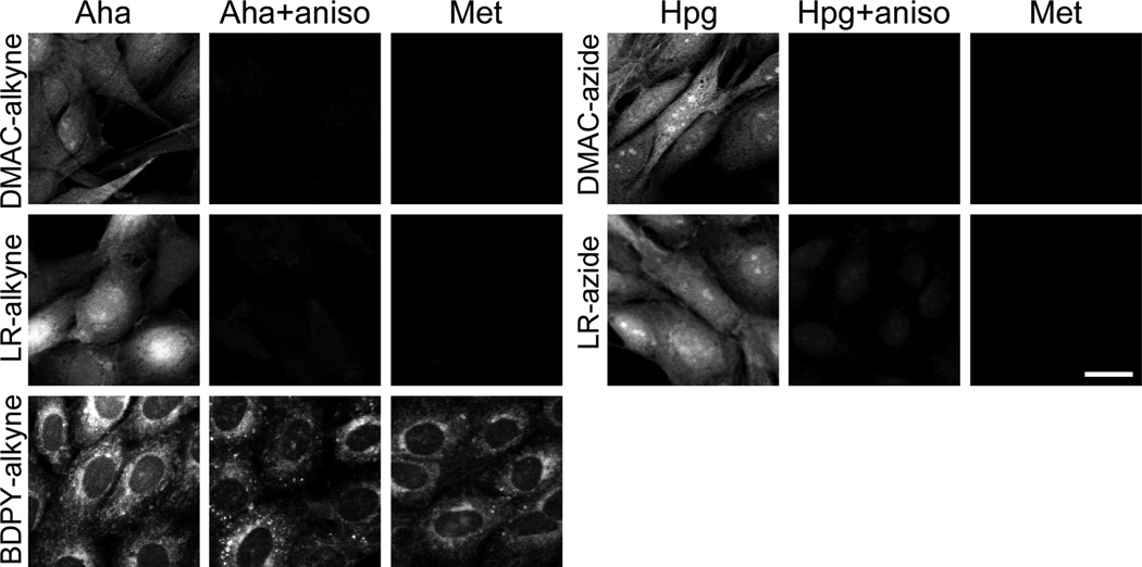
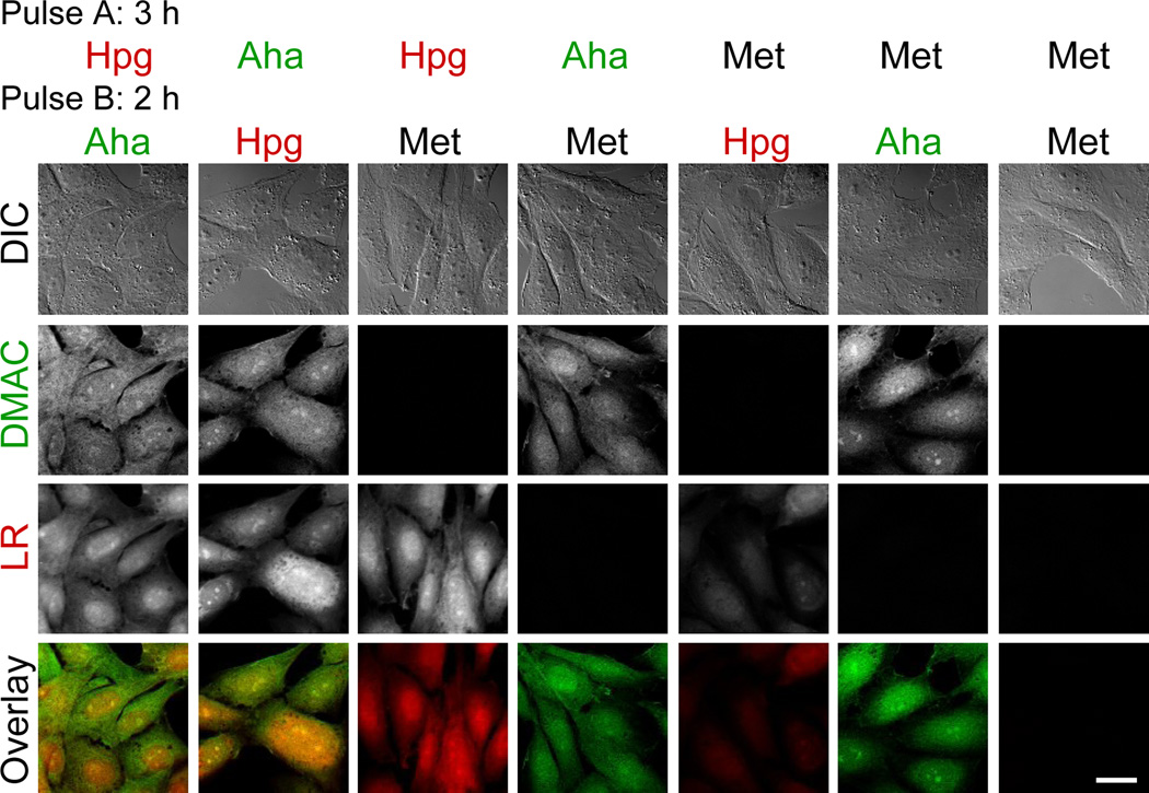
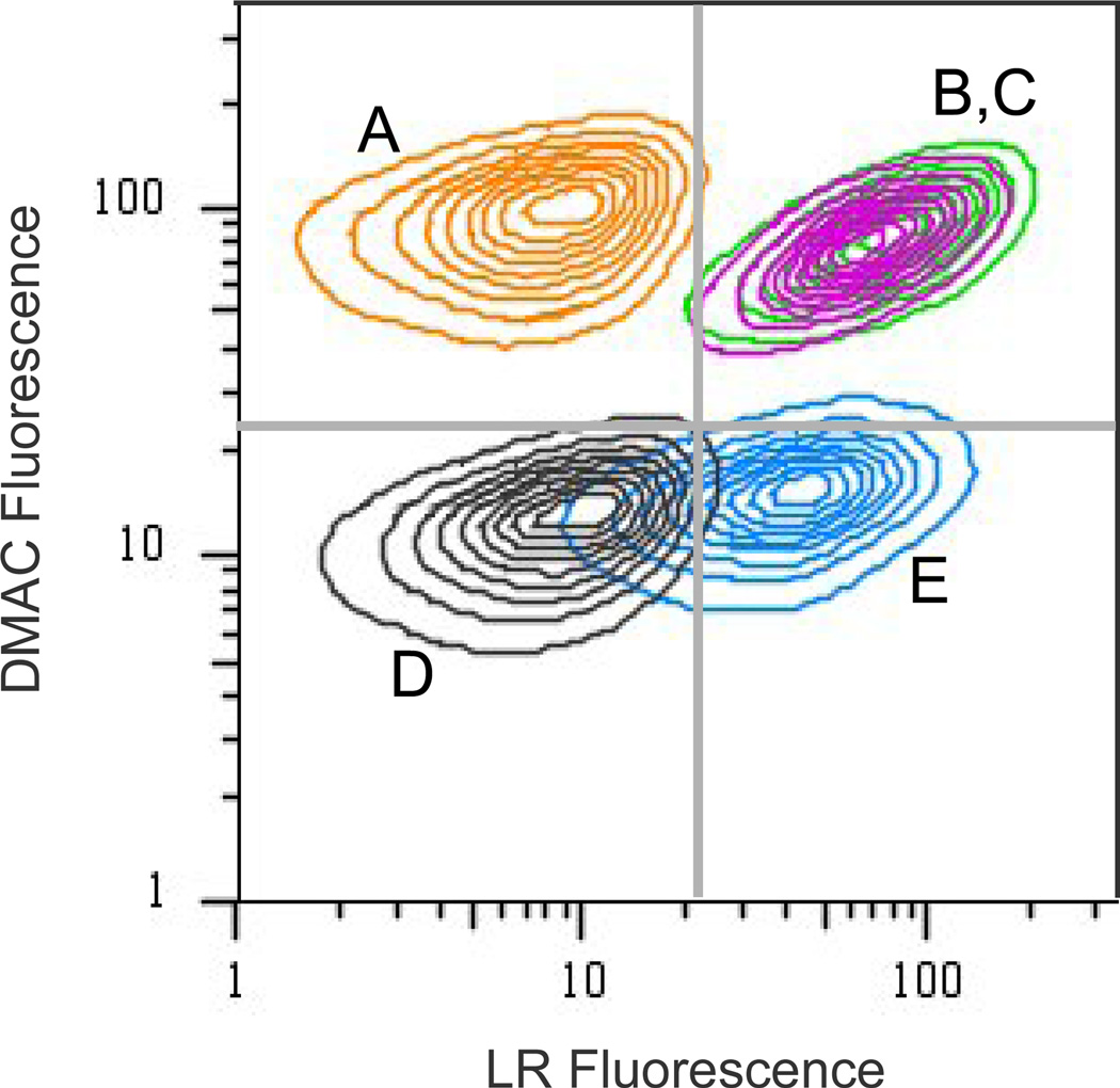
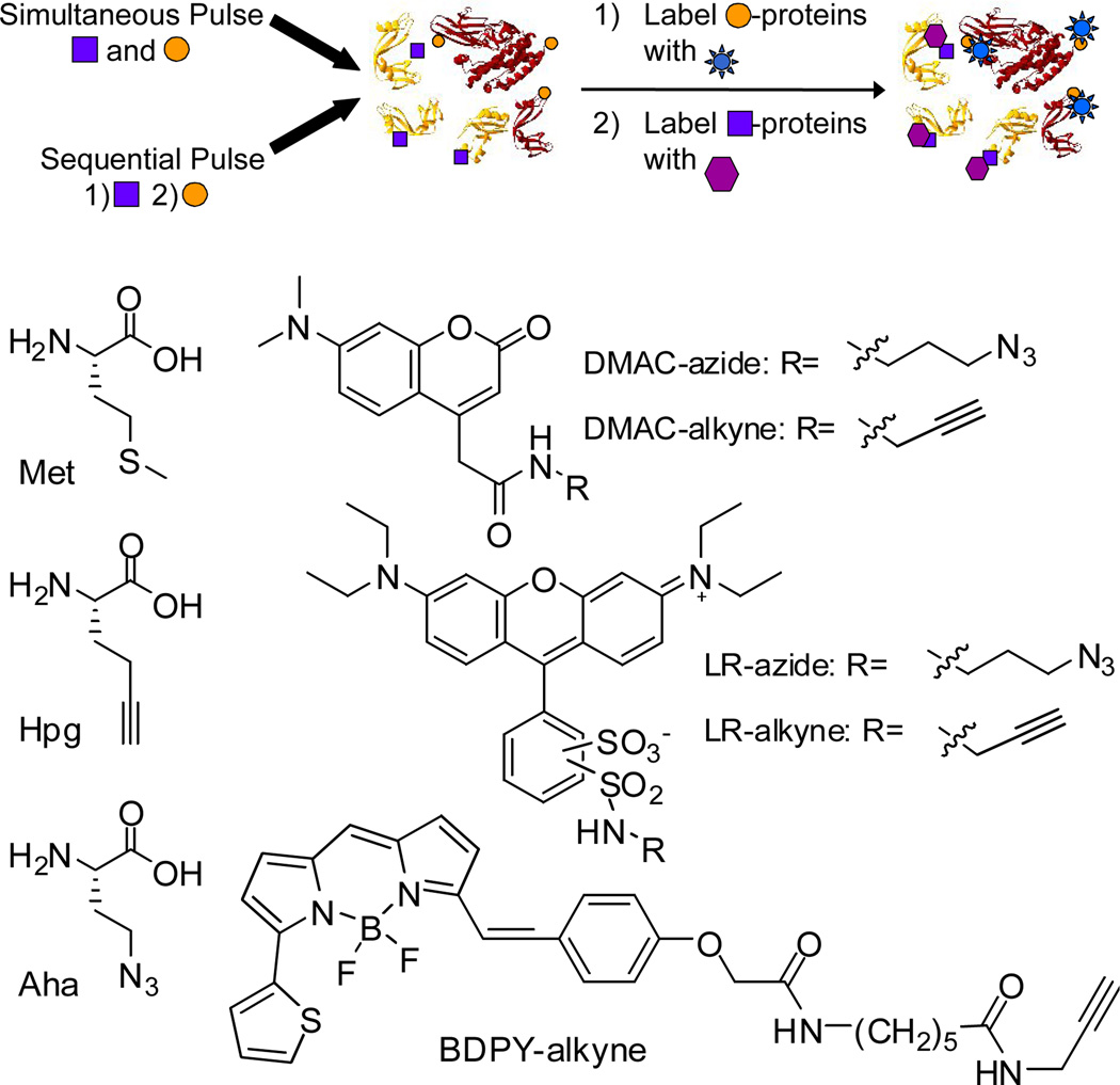
The proteome undergoes complex changes in response to disease, drug treatment, and normal cellular signaling processes. Characterization of such changes requires methods for time-resolved protein identification and imaging. Here, we describe the application of two reactive methionine (Met) analogues, azidohomoalanine (Aha) and homopropargylglycine (Hpg), to label two protein populations in fixed cells. Reactive lissamine rhodamine (LR), 7-dimethylaminocoumarin (DMAC), and bodipy-630 (BDPY) dyes were prepared and examined for use in selective dye-labeling of newly synthesized proteins in Rat-1 fibroblasts. The LR and DMAC, but not BDPY, fluorophores were found to enable selective, efficient labeling of subsets of the proteome; cells labeled with Aha and Hpg exhibited fluorescence emission three- to sevenfold more intense than that of control cells treated with Met. We also examined simultaneous and sequential pulse-labeling of cells with Aha and Hpg. After pulse-labeling, cells were treated with reactive LR and DMAC dyes, and labeled cells were imaged by fluorescence microscopy and analyzed by flow cytometry. The results of these studies demonstrate that amino acid labeling can be used to achieve selective two-color imaging of temporally defined protein populations in mammalian cells.


Figures

Figure 1
Figure 1
Selective dye-labeling of newly synthesized proteins using azide or alkyne fluorophores. Confocal fluorescence imaging of Rat-1 fibroblasts grown for 3 h in media containing 1 mM Met, 1 mM Aha, or 1 mM Hpg. Control cells were pre-treated with the protein synthesis inhibitor anisomycin (aniso) prior to pulse-labeling. Cells were dye labeled with 10 µM DMAC-alkyne, 50 µM LR-alkyne, 10 µM BDPY-alkyne, 50 µM DMAC-azide, or 50 µM LR-azide. Scale bar represents 20 µm. Corresponding DIC images can be found in the Supporting Information (Figure S1).
Figure 2
Figure 2
Fluorescent images of Rat-1 fibroblasts simultaneously pulse-labeled with two reactive amino acids. Cells were pulse-labeled for 5 h with 1 mM Met, 1 mM Aha and 1 mM Hpg (1:1), 3 mM Aha and 1 mM Hpg (3:1), or 1 mM Aha and 3 mM Hpg (1:3). Cells were fixed and blocked before dye-labeling with LR-azide and DMAC-alkyne. Images were false-colored in ImageJ. Scale bar represents 20 µm.
Figure 3
Figure 3
Fluorophore labeling of two distinct populations of proteins in Rat-1 fibroblasts. Cell were pulse-labeled for 3 h with an amino acid (Pulse A), washed, and then pulse-labeled for 2 h with a second amino acid (Pulse B). Cells were fixed and blocked before dye-labeling for 1 h with 50 µM LR-azide and 1 h with 10 µM DMAC-alkyne. The overlay shows both the DMAC (green) and LR (red) fluorescence. Scale bar represents 20 µm.
Figure 4
Figure 4
Flow cytometry contour plot of two-dye labeled fibroblasts. Cell were pulse-labeled for 3 h with 1 mM amino acid (Pulse I), washed, and then pulse-labeled for 2 h with 1 mM of a second amino acid (Pulse II). Cells were fixed and blocked before dye-labeling with LR-azide and DMAC-alkyne. A: Cells were pulse-labeled with 1 mM Aha, followed by 1 mM Met [Orange: Aha→Met]. B: [Magenta: Aha→Hpg]. C: [Green: Hpg→Aha]. D: [Grey: Met→Met]. E: [Blue: Hpg→Met]. For each sample, 50,000 total events were collected. Dead cells and debris were excluded from analysis using forward-scatter and side-scatter.
Scheme 1
Scheme 1
Two-dye labeling strategy and structures of Met, Hpg, Aha, and the reactive fluorophores.

References

    1. Lemieux GA, Bertozzi CR. Trends Biotechnol. 1998;16:506. - PubMed
    1. Rodriguez EC, Marcaurelle LA, Bertozzi CR. J. Org. Chem. 1998;63:7134. - PubMed
    1. Datta D, Wang P, Carrico IS, Mayo SL, Tirrell DA. J. Am. Chem. Soc. 2002;124:5652. - PubMed
    1. Wang L, Zhang ZW, Brock A, Schultz PG. Proc. Natl. Acad. Sci. USA. 2003;100:56. - PMC - PubMed
    1. Kiick KL, Saxon E, Tirrell DA, Bertozzi CR. Proc. Natl. Acad. Sci. USA. 2002;99:19. - PMC - PubMed

Publication types