. 2008 Sep 5;134(5):757-68.
doi: 10.1016/j.cell.2008.07.030.

Crossing the species barrier by PrP(Sc) replication in vitro generates unique infectious prions

Affiliations

Crossing the species barrier by PrP(Sc) replication in vitro generates unique infectious prions

Joaquín Castilla et al. Cell. .

Abstract

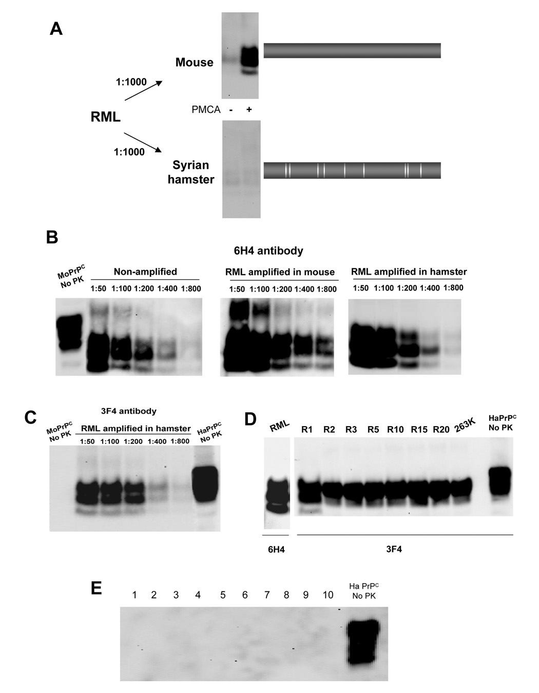
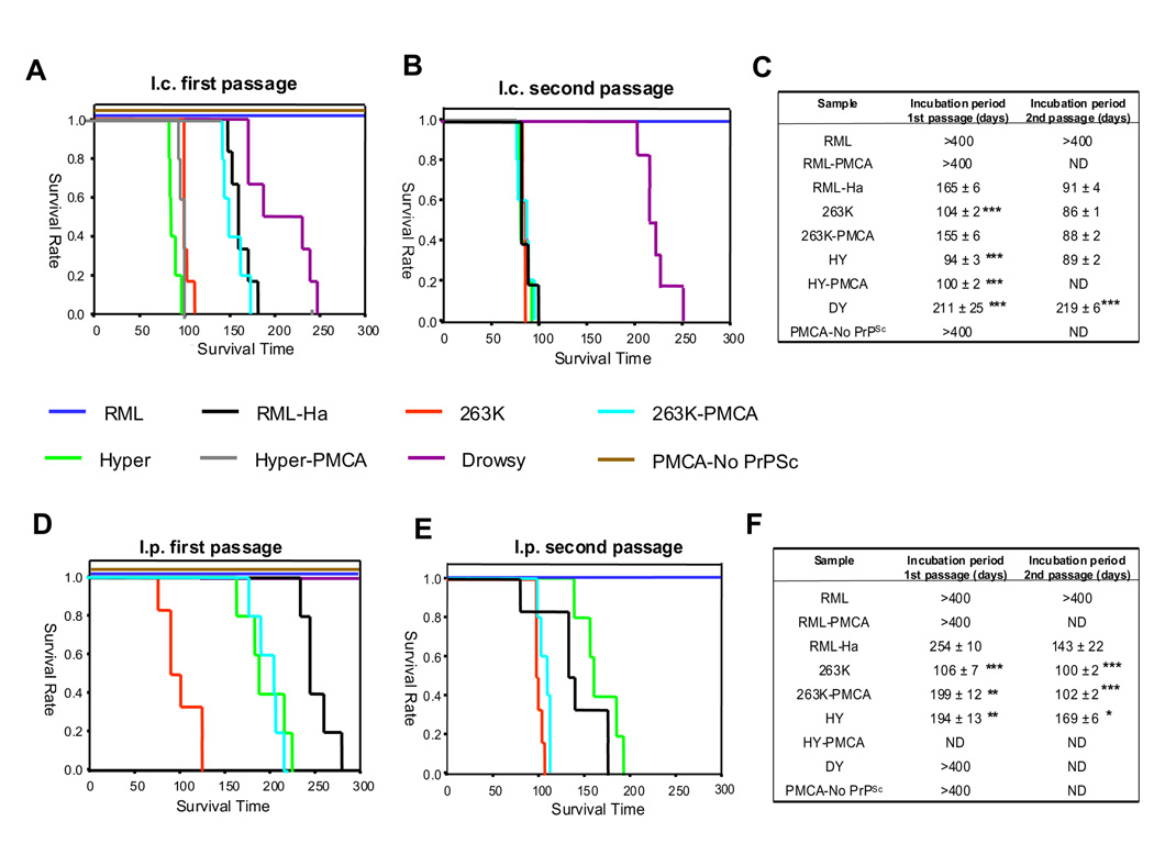
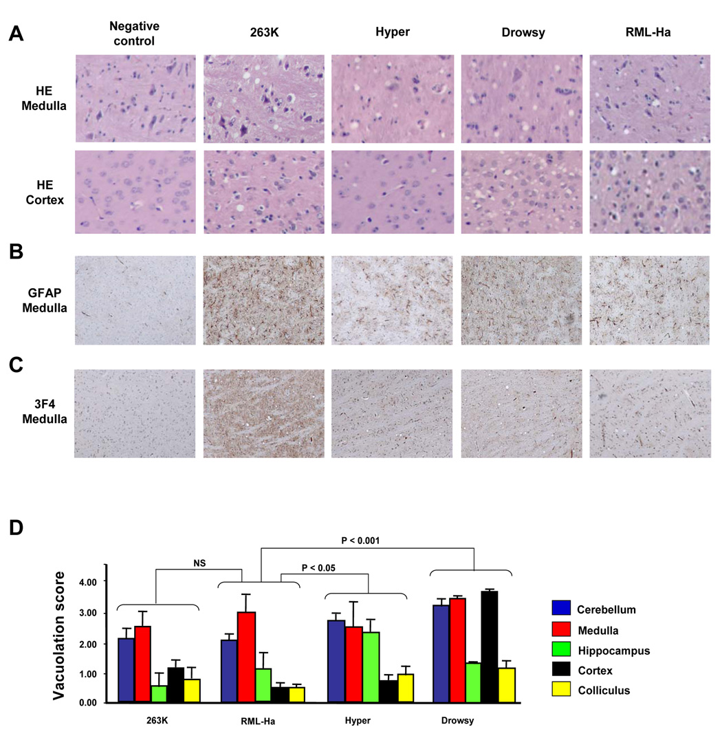
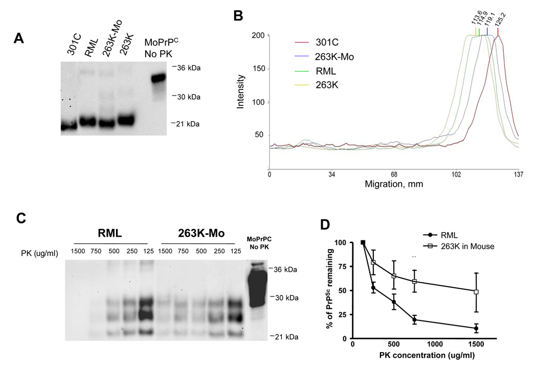
Prions are unconventional infectious agents composed exclusively of misfolded prion protein (PrP(Sc)), which transmits the disease by propagating its abnormal conformation to the cellular prion protein (PrP(C)). A key characteristic of prions is their species barrier, by which prions from one species can only infect a limited number of other species. Here, we report the generation of infectious prions by interspecies transmission of PrP(Sc) misfolding by in vitro PMCA amplification. Hamster PrP(C) misfolded by mixing with mouse PrP(Sc) generated unique prions that were infectious to wild-type hamsters, and similar results were obtained in the opposite direction. Successive rounds of PMCA amplification result in adaptation of the in vitro-produced prions, in a process reminiscent of strain stabilization observed upon serial passage in vivo. Our results indicate that PMCA is a valuable tool for the investigation of cross-species transmission and suggest that species barrier and strain generation are determined by the propagation of PrP misfolding.


Figures

Figure 1
Figure 1. In vitro conversion of hamster PrPC induced by mouse RML PrPSc
A: RML brain homogenate was diluted 1000-fold into either mouse or hamster normal brain homogenate and subjected to 96 PMCA cycles. The blot shows the results with and without PMCA in each species. At the right side we show a scheme of PrP indicating the position in which there are amino acid differences between mice and hamsters. B: To attempt forcing conversion, larger quantities (dilutions 1:50 thru 1:800) of RML PrPSc were incubated with mouse (central panel) or hamster (right panel) PrPC. All samples (except for the control samples in the left panel labeled as “non-amplified”) were subjected to 96 PMCA cycles and PrPSc signal was detected after PK digestion by western blot using the 6H4 antibody. C: The same samples as in the right panel of B were developed using the 3F4 antibody. D: The newly generated RML-Ha PrPSc was serially passed in hamster brain homogenate by a series of 1:10 dilution following by 48 PMCA cycles. R indicates the number of rounds of PMCA, i.e. R5 represent the samples after 5 serial rounds of PMCA. E: To assess spontaneous generation of PrPSc by PMCA, samples from brain of 10 different hamsters were subjected to the same process of serial PMCA as in panel D. PrPSc formation was analyzed by western blot after PK treatment in each PMCA round. The figure shows the results obtained after 20 rounds of PMCA. In the experiments shown in this figure all samples were treated with PK, except when indicated.
Figure 2
Figure 2. Infectivity of newly generated RML-Ha PrPSc after crossing the species barrier
RML-Ha PrPSc samples amplified by 20 serial PMCA rounds were inoculated i.c. or i.p. into 6 wild type hamsters. As controls we inoculated similar quantities of PrPSc from RML or three distinct hamster strains (263K, Hyper and Drowsy). We also show the data obtained by inoculation of in vitro generated prions through 20 serial rounds of PMCA by incubation of 263K (263K-PMCA) or Hyper (HY-PMCA) PrPSc with healthy hamster brain homogenate and RML replicated at expenses of mouse PrPC (RML-PMCA). The figure also show the results obtained by inoculation of the material produced after 20 rounds of PMCA using unseeded normal hamster brain homogenate (PMCA-No PrPSc). Panels A and D show the survival curves obtained after i.c. and i.p. inoculation, respectively, of the in vitro generated RML-Ha after 20 rounds of PMCA. Panels B and E show the survival curves of the second passage (i.e. animals were inoculated with material obtained from the brain of sick animals in the experiments depicted in panels A and D) after i.c and i.p inoculation, respectively. Panels C and F show the average incubation periods of the experiments done by i.c. and i.p. inoculation of various samples. The values correspond to the average ± standard error. The data was analyzed by ANOVA and the Dunnett multiple comparison post-test. Each set of data was compared to the results obtained with the RML-Ha strain and significant differences are highlighted with asterisks (* = P<0.05; ** = P<0.01; *** = P<0.001).
Figure 3
Figure 3. Histopathological features of the disease induced by inoculation of hamsters with PMCA-generated RML-Ha PrPSc
Brain from sick animals in which disease was produced by inoculation with the in vitro generated RML-Ha PrPSc (first passage) or the known hamster strains 263K, Hyper and Drowsy were analyzed by histological studies. As control we used the brain of a hamster inoculated with PBS and sacrificed without disease at 350 days post-inoculation. A: Spongiform degeneration was evaluated after haematoxilin-eosin staining of medulla and occipital cortex sections, and visualized by microscopy at a 40X magnification. B: Reactive astroglyosis was evaluated by histological staining with glial fibrillary acidic protein antibody. C: PrP accumulation in these animals was evaluated by staining the tissue with the 3F4 anti-PrP monoclonal antibody. D: The vacuolation profile in each brain area was estimated using a semi-quantitative scale, as described in Experimental procedures. The brain areas used were the following: occipital cortex, cerebellum (mostly white matter), medulla (spinal 5 nucleus, interpolar part), inferior colliculum and hippocampus (CA1 and CA2 regions). We also included in the analysis brain sections from animals inoculated with the other hamster prion strains. The values represent the average ± standard error of the extent of vacuolation from the 5 animals analyzed in each set. Statistical analysis by two-ways ANOVA, using brain regions and prion origin as the variables indicated that differences were highly significant (P<0.001). To assess the significance of the differences between each known prion strain and RML-Ha, we used the Dunnett multiple comparison post-test and the P values for each combination are shown in the figure.
Figure 4
Figure 4. Biochemical characterization of RML-Ha PrPSc
Samples from brains of animals inoculated with RML-Ha PrPSc (first passage in vivo) were used to study the PK resistance profile (A), the relative mobility after deglycosylation and PK treatment (B) and the susceptibility to guanidine denaturation (C). As controls, we used samples from RML or three distinct hamster strains (263K, Hyper and Drowsy). The results in panel A and C correspond to the quantitative evaluation of western blots by densitometric analysis from 3 independent animals. The data represent the average ± standard error. The data was analyzed by ANOVA and the Dunnett multiple comparison post-test. Each set of data was compared to the results obtained with the RML-Ha strain and significant differences are highlighted with asterisks (* = P<0.05; ** = P<0.01; *** = P<0.001).
Figure 5
Figure 5. In vitro conversion of mouse PrPC induced by hamster 263K PrPSc generates infectious prions
A: Schematic representation of the dilutions done and the PMCA rounds used for our in vivo infectivity experiments. B: Survival curve observed after inoculation of 6 wild type mice with the material generated after several rounds of PMCA. R indicates the number of rounds of PMCA. As control the animals were inoculated with 263K hamster prions. C: Average and standard error of the incubation times and attack rates observed after inoculation of wild type mice with the material produced after different rounds of PMCA. D: Comparison of survival curves for the stabilized 263K-Mo infectious material (after 15 rounds of PMCA) with those obtained with RML and 301C, two mouse strains of different origin. We also show the data obtained by inoculation of in vitro generated prions through 20 serial rounds of PMCA by incubation of RML (RML-PMCA) or 301C (301C-PMCA) PrPSc with healthy mouse brain homogenate. The figure also show the results obtained by inoculation of the material produced after 20 rounds of PMCA using unseeded normal mouse brain homogenate (PMCA-No PrPSc), which correspond to the control for de novo generation of prions. For all these experiments the material was inoculated i.c. as described in Experimental results.
Figure 6
Figure 6. Histopathological features of the disease induced by inoculation of mice with PMCA-generated 263K-Mo PrPSc
Brain from sick mice in which disease was produced by inoculation with the newly generated 263K-Mo prions after 15 rounds of PMCA (first passage) or the known mouse strains RML and 301C were analyzed by histological studies. As control we used brain of a mouse inoculated with PBS and sacrificed without disease at 350 days post-inoculation. A: Spongiform degeneration was evaluated after haematoxilin-eosin (HE) staining of three different brain areas (cerebellum, medulla and hippocampus) and was visualized at a 40X magnification. B: Reactive astroglyosis was evaluated in the inferior culliculus by staining with glial fibrillary acidic protein antibody. C: PrP accumulation in these animals was evaluated in the occipital cortex and cerebellum by staining the tissue with the 6H4 antibody. D: The vacuolation profile in each brain area was estimated using a semi-quantitative scale, as described in Experimental procedures. The brain areas used were the following: occipital cortex, cerebellum (mostly white matter), medulla (spinal 5 nucleus, interpolar part), inferior colliculum and hippocampus (CA1 and CA2 regions). We also included in the analysis brain sections from animals inoculated with RML and 301C. The values represent the average ± standard error of the extent of vacuolation from the 5 animals analyzed in each set. Statistical analysis by two-ways ANOVA, using brain regions and prion origin as the variables indicated that differences were highly significant (P<0.001). To assess the significance of the differences between each known prion strain and 263K-Mo, we used the Dunnett multiple comparison post-test and the P values for each combination are shown in the figure.
Figure 7
Figure 7. Biochemical characteristcs of 263K-Mo PrPSc
A: Samples from brains of mice inoculated with 263K-Mo, RML or 301C were used to study the electrophoretical migration after deglycosylation and PK treatment. B: To assess the electrophoretical differences among distinct strains, the blot in panel A was scanned and analyzed by a software included in the UVp image analysis system to locate the exact position of the bands. C: The PK resistance profile of 263K-Mo PrPSc was studied and compared with RML. D: The results of the experiment shown in panel B was quantitated by densitometric analysis. The data in the figure represent the average ± standard error from 3 independent animals. The differences were statistically significant as evaluated by one-way ANOVA (P<0.01).

References

    1. Atarashi R, Moore RA, Sim VL, Hughson AG, Dorward DW, Onwubiko HA, Priola SA, Caughey B. Ultrasensitive detection of scrapie prion protein using seeded conversion of recombinant prion protein. Nat. Methods. 2007;4:645–650. - PubMed
    1. Brachmann A, Baxa U, Wickner RB. Prion generation in vitro: amyloid of Ure2p is infectious. EMBO J. 2005;24:3082–3092. - PMC - PubMed
    1. Bruce ME. 'New variant' Creutzfeldt-Jakob disease and bovine spongiform encephalopathy. Nat. Med. 2000;6:258–259. - PubMed
    1. Bruce ME. TSE strain variation. Br. Med. Bull. 2003;66:99–108. - PubMed
    1. Buschmann A, Groschup MH. TSE eradication in small ruminants--quo vadis? Berl Munch. Tierarztl. Wochenschr. 2005;118:365–371. - PubMed

Publication types