. 2008 Sep 5;134(5):769-81.
doi: 10.1016/j.cell.2008.06.037.

Chemical and biological approaches synergize to ameliorate protein-folding diseases

Affiliations

Chemical and biological approaches synergize to ameliorate protein-folding diseases

Ting-Wei Mu et al. Cell. .

Abstract

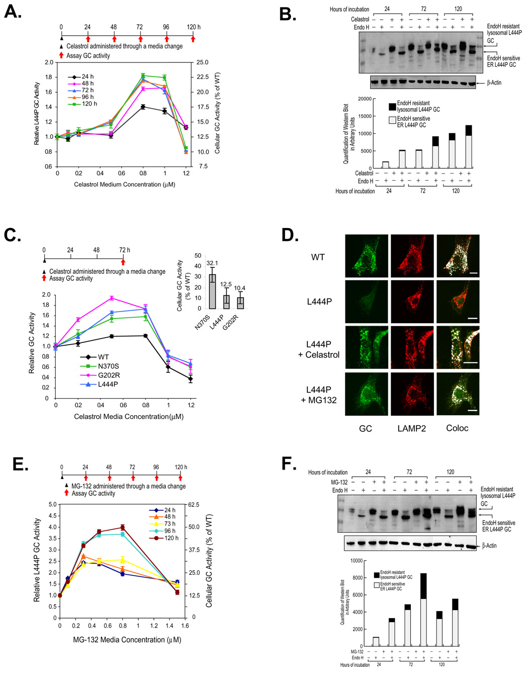
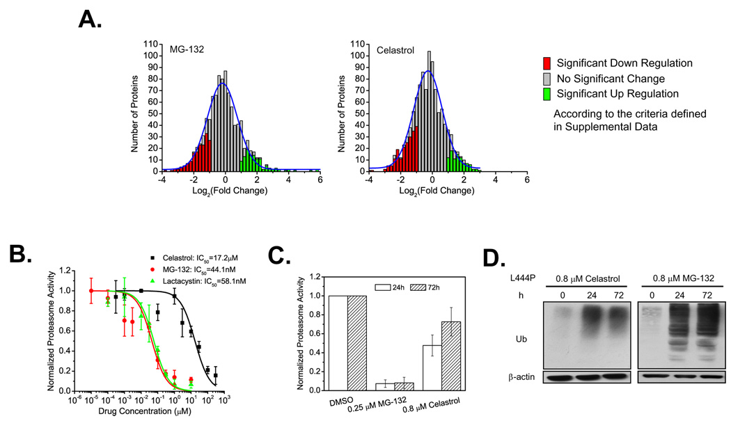
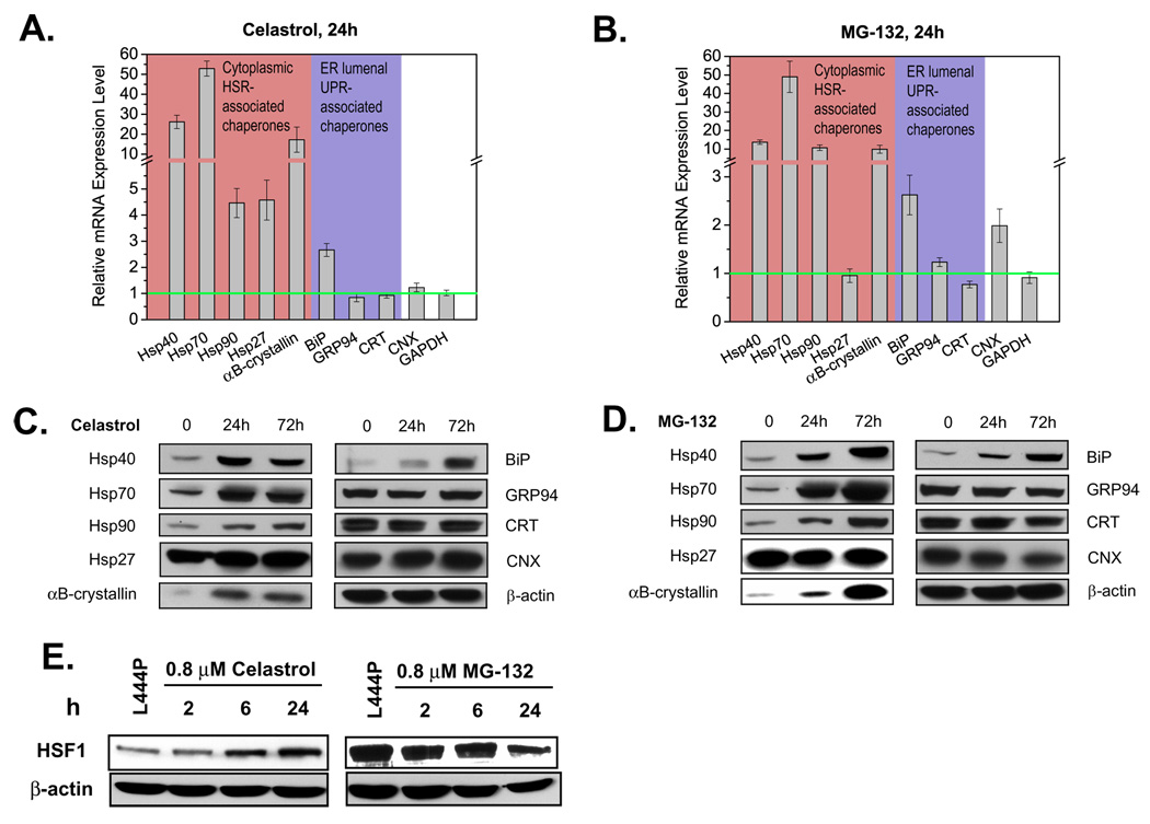
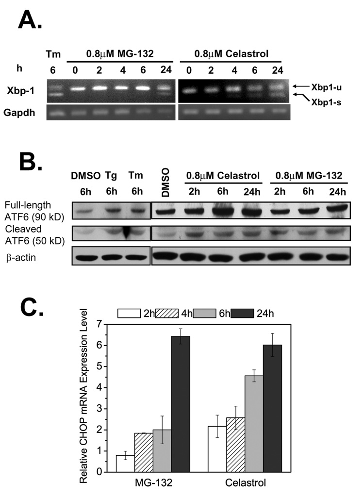
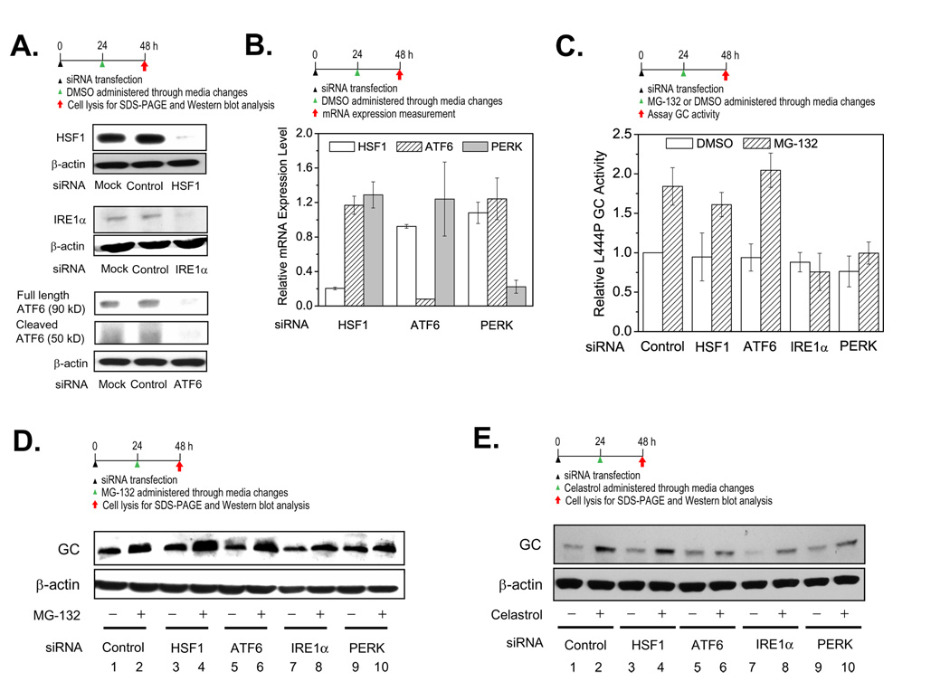
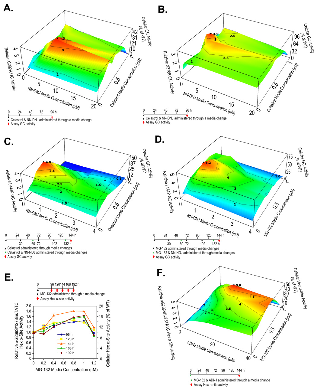
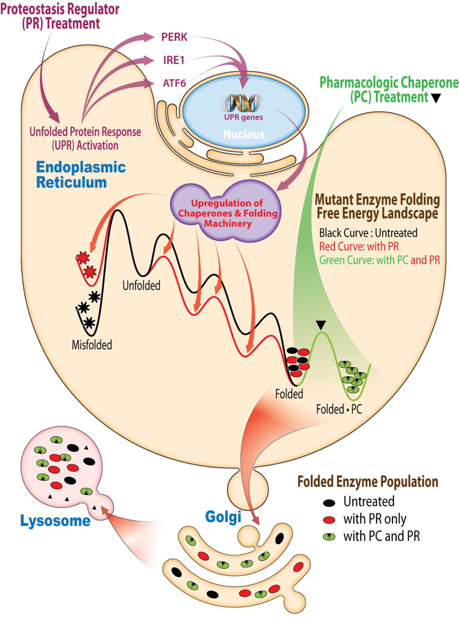
Loss-of-function diseases are often caused by a mutation in a protein traversing the secretory pathway that compromises the normal balance between protein folding, trafficking, and degradation. We demonstrate that the innate cellular protein homeostasis, or proteostasis, capacity can be enhanced to fold mutated enzymes that would otherwise misfold and be degraded, using small molecule proteostasis regulators. Two proteostasis regulators are reported that alter the composition of the proteostasis network in the endoplasmic reticulum through the unfolded protein response, increasing the mutant folded protein concentration that can engage the trafficking machinery, restoring function to two nonhomologous mutant enzymes associated with distinct lysosomal storage diseases. Coapplication of a pharmacologic chaperone and a proteostasis regulator exhibits synergy because of the former's ability to further increase the concentration of trafficking-competent mutant folded enzymes. It may be possible to ameliorate loss-of-function diseases by using proteostasis regulators alone or in combination with a pharmacologic chaperone.


Figures

Figure 1
Figure 1
Celastrol or MG-132 treatment enhances folding, trafficking and activity of glucocerebrosidases (GCs) variants. Relative lysosomal GC activity of L444P GC fibroblasts upon celastrol (A) or MG-132 (E) treatment. Reported activities were normalized to the activity of untreated cells of the same type (left y axis) and expressed as the percentage of WT GC activity (right y axis). Western blot analysis of L444P GC trafficking within fibroblasts after 24, 72, and 120 h exposure to 0.8 µM celastrol (B) or 0.8 µM MG-132 (F). The white portion of the bars represents quantification (Java Image processing and analysis software from the NIH) of the lower, endo H sensitive bands and the black portion of the bars represents the higher MW, endo H resistant post-Golgi glycoform. Relative lysosomal activity of WT GC and Gaucher disease-associated N370S, G202R, and L444P GC variants (C) in patient-derived fibroblasts. The inset displays GC variant enzyme activity expressed as a percentage of WT GC activity, as reported previously (Sawkar et al., 2005). The data in (A), (C) and (E) were reported as mean ± SEM. Immunofluorescence microscopy of GC (D) in L444P fibroblasts and WT cells (positive control). L444P GC cells were untreated (second row) or incubated with 0.8 µM celastrol (third row) or 0.25 µM MG-132 (bottom row) for 3 days. GC was detected using the mouse anti-GC antibody 8E4 (column 1); rabbit anti-LAMP2 antibody was used as a lysosomal marker (column 2). Colocalization of GC (green) and LAMP2 (red) was artificially colored white (column 3). Bar equals 20 µM.
Figure 2
Figure 2
Influence of celastrol and MG-132 on proteostasis. Changes in the L444P GC fibroblast proteome (A) after MG-132 (0.8 µM) or celastrol (0.8 µM) treatment for 72 h. The number of proteins is plotted against fold change using a normalized spectra count ratio of drug treated samples versus untreated samples in cases where a given protein is detected in both untreated and treated samples. Dose-response curves (B) of the inhibition of the chymotrypsin-like activity of the proteasome by celastrol, MG-132, or lactacystin in L444P GC cells. Chymotrypsin-like activity of the proteasome (C) in L444P GC cells incubated with celastrol (0.8 µM) or MG-132 (0.25 µM) for 24 h and 72 h. The data were reported as mean ± SD in (B) or as mean ± SEM in (C). Western blot analysis of ubiquitinated proteome (D) in L444P GC fibroblasts after 0.8 µM celastrol or MG-132 treatment for 24 h and 72 h.
Figure 3
Figure 3
Both MG-132 and celastrol activate the HSR in L444P GC fibroblasts. Relative chaperone mRNA expression probed by quantitative RT-PCR in cells treated with 0.8 µM celastrol (A) or 0.8 µM MG-132 (B) for 24 h. Relative mRNA expression level for treated L444P GC cells normalized to that of untreated cells after correction for the expression level of GAPDH, a housekeeping control. The data in (A) and (B) were reported as mean ± SEM. Western blot analyses in L444P GC fibroblasts treated with 0.8 µM celastrol (C) or 0.8 µM MG-132 (D) for 24 h or 72 h. HSF1 protein expression levels (E) in L444P GC cells treated with 0.8 µM celastrol and 0.8 µM MG-132.
Figure 4
Figure 4
Activation of the UPR by MG-132 and celastrol. Detection of spliced Xbp-1 mRNA by RT-PCR (A) in L444P GC fibroblasts treated with 0.8 µM MG-132 or 0.8 µM celastrol (tunicamycin (Tm) is a positive control, GAPDH, a housekeeping control). Xbp1-u represents unspliced Xbp-1, and Xbp1-s represents spliced Xbp-1. Cleavage of ATF6 proteins revealed by Western blotting (B) in celastrol or MG-132 treated L444P GC cells (thapsigargain (Tg) and Tm served as positive controls). Relative mRNA expression levels of CHOP (C) probed by quantitative RT-PCR in L444P GC fibroblasts treated with 0.8 µM celastrol or MG-132. Relative mRNA expression level for treated L444P GC cells was normalized to that of untreated cells after correction for the expression level of GAPDH. The data in (C) were reported as mean ± SEM.
Figure 5
Figure 5
The UPR, but not the HSR, is important for PR function. Knockdown of HSF1, IRE1α, and ATF6 (A) by siRNA lasts at least 48 h, according to Western blot analysis. Relative mRNA expression levels of HSF1, ATF6 and PERK were probed by quantitative RT-PCR to verify knockdown of HSF1, ATF6 and PERK (B) in L444P GC fibroblasts after specific siRNA treatment—mRNA expression levels normalized to that of non-targeting siRNA treated cells after correction for GAPDH levels. siRNA knockdown of IRE1α or PERK (C) blocks the ability of MG-132 (0.25 µM in DMSO) to increase L444P GC activity, activities normalized to L444P GC cells treated with both non-targeting siRNA (control) and DMSO vehicle. The data in (B) and (C) were reported as mean ± SD. Western blot analysis of L444P GC in fibroblasts treated with non-targeting siRNA (control) plus DMSO (vehicle) or HSF1, IRE1α, ATF6 and PERK siRNAs without (just DMSO vehicle) or with 0.25 µM MG-132 (D) or 0.8 µM celastrol (E) in DMSO.
Figure 6
Figure 6
PCs and PRs exhibit synergy in Gaucher and Tay-Sachs fibroblasts. Enzyme activities within patient-derived fibroblasts treated with a PC and a PR. In all the 3D plots (A–D, and F), PR and PC media concentrations are shown on the x and y-axes, and the mutant enzyme activities on the z-axis with the dosing schematic depicted at the bottom. Reported activities were normalized to the activity of untreated cells of the same type (left z axis) and expressed as the percentage of WT enzyme activity (right z axis). αG269S/1278insTATC HexA fibroblasts were exposed to MG-132 (E) and the Hex α-site activities were measured as a function of time. The data in (E) were reported as mean ± SEM.
Figure 7
Figure 7
PRs and PCs synergize to restore mutant enzyme homeostasis. PRs activate the UPR, resulting in the coordinated transcription and translation of chaperones and folding enzymes that resculpt the folding free energy diagram of the mutant enzyme in the ER, enhancing its folding and minimizing misfolding, which increases the population of the mutant enzyme in the folded state. PCs bind to the folded state of the mutant enzyme, further stabilizing it, which further increases its population. Together these mechanisms of action are more effective at increasing the concentration of the folded mutant enzyme and folded mutant enzyme•PC complex, both of which can engage the trafficking machinery, increasing mutant enzyme lysosomal concentration and activity.

References

    1. Albanese V, Yam AY-W, Baughman J, Parnot C, Frydman J. Systems analyses reveal two chaperone networks with distinct functions in eukaryotic cells. Cell. 2006;124:75–88. - PubMed
    1. Awasthi N, Wagner BJ. Upregulation of heat shock protein expression by proteasome inhibition: An antiapoptotic mechanism in the lens. Invest Ophthalmol Vis Sci. 2005;46:2082–2091. - PubMed
    1. Balch WE, Morimoto RI, Dillin A, Kelly JW. Adapting proteostasis for disease intervention. Science. 2008;319:916–919. - PubMed
    1. Beutler E. Lysosomal storage diseases: natural history and ethical and economic aspects. Mol Genet Metab. 2006;88:208–215. - PubMed
    1. Beutler E, Dale GL, Guinto E, Kuhl W. Enzyme replacement therapy in Gaucher's disease: Preliminary clinical trial of a new enzyme preparation. Proc Natl Acad Sci USA. 1977;74:4620–4623. - PMC - PubMed

Publication types