. 2008 Sep 5;134(5):843-53.
doi: 10.1016/j.cell.2008.06.053.

In vivo imaging of oskar mRNA transport reveals the mechanism of posterior localization

Affiliations

In vivo imaging of oskar mRNA transport reveals the mechanism of posterior localization

Vitaly L Zimyanin et al. Cell. .

Abstract

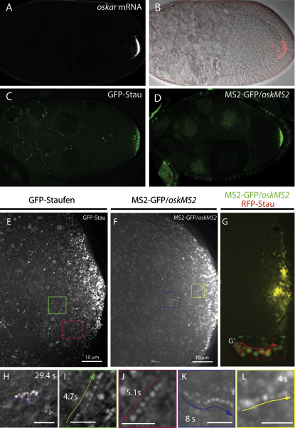
oskar mRNA localization to the posterior of the Drosophila oocyte defines where the abdomen and germ cells form in the embryo. Although this localization requires microtubules and the plus end-directed motor, kinesin, its mechanism is controversial and has been proposed to involve active transport to the posterior, diffusion and trapping, or exclusion from the anterior and lateral cortex. By following oskar mRNA particles in living oocytes, we show that the mRNA is actively transported along microtubules in all directions, with a slight bias toward the posterior. This bias is sufficient to localize the mRNA and is reversed in mago, barentsz, and Tropomyosin II mutants, which mislocalize the mRNA anteriorly. Since almost all transport is mediated by kinesin, oskar mRNA localizes by a biased random walk along a weakly polarized cytoskeleton. We also show that each component of the oskar mRNA complex plays a distinct role in particle formation and transport.


Figures

Figure 1
Figure 1
osk mRNA Particles Undergo Fast, Directed Movements in All Directions in the Oocyte Cytoplasm (A and B) osk mRNA localization to the posterior of a wild-type stage 9 oocyte. (B) An overlay of a pseudo differential interference contrast image and the in situ hybridization signal (red channel) from (A). (C) GFP-Stau localization in a live stage 9 oocyte. (D) The localization of osk mRNA labeled with MS2-GFP in a live stage 9 egg chamber. MS2-GFP contains a nuclear localization signal and therefore localizes to the nurse cell nuclei when not associated with osk mRNA. (E and F) Overlays of 25 frames from timelapse movies of wild-type stage 9 oocytes expressing GFP-Stau (E) or oskMS2/MS2-GFP (F) to show the movements of osk mRNA particles. Examples of individual tracks are highlighted by colored rectangles and shown in (I)–(L). (G) oskMS2/MS2-GFP and RFP-Stau colocalize at the posterior pole of the oocyte and in individual moving particles. (G′) shows an overlay of 25 frames from a movie in which the red and green channels were imaged alternately. See also Movie S5. (H) An example of a particle moving passively with the cytoplasmic flows and undergoing Brownian motion. (I–L) Closeups of the fast, directed particle tracks, highlighted by the colored rectangles in (G) and (H). GFP-Stau (I and J); oskMS2/MS2-GFP (K and L). Scale bars are 2 μm.
Figure 2
Figure 2
osk mRNA Particle Movements Show a Weak Posterior Bias (A) A graph showing the average speeds of GFP-Stau and oskMS2/MS2-GFP particle movements in the oocyte and in the anterior and posterior halves of the oocyte. The error bars show the standard error of the mean (SEM). (B) The distribution of GFP-Stau particle speeds in the anterior (black) and posterior (red) halves of the oocyte. (C) Circular graphs showing the orientation of GFP-Stau (i and ii) and oskMS2/MS2-GFP (iii and iv) particle tracks. Both methods reveal a significant posterior bias in the direction of particle movements (p < 0.005 and p < 0.025, χ2 test). (D) The posterior bias of particle movement results in an overall positive net posterior displacement of GFP-Stau and oskMS2/MS2-GFP particles in the oocyte. Error bars show SEM.
Figure 3
Figure 3
Behavior of osk RNP Particles in Localization Mutants (A–F″) Low magnification images (A–F) and overlays of 25 frames from high-magnification timelapse movies of oskMS2 (A and F) or GFP-Stau (B–E). The blue lines indicate the nurse cell/oocyte boundary. The posterior pole of the oocyte is marked with white asterisks. (A–A″) Wild-type. (A″) shows a closeup of the track marked by the red rectangle in (A′). (B–B″) hrp4810B2-9 germline clones. There are no detectable particles in this mutant. (C–C″) btz2 germline clones. (D–D″) mago1/Df(2R)F36. (E–E″) TmIIgs. (F–F″) stauD3. (G) A graph showing the proportion of particles that undergo fast, directed movements in a normalized area of cytoplasm in wild-type, stau, and mago mutant oocytes. Error bars show SEM. (H) A bar chart showing the percentages of fast particle movements toward the anterior and posterior of the oocyte in wild-type, mago, btz2, TmIIgs, and stau mutants. The asterisks indicate the mutants in which the bias differs significantly from wild-type. (χ2 test: p ≤ 0.05; ∗∗p ≤ 0.01; ∗∗∗p ≤ 0.001.) (I) A bar chart showing the net posterior displacement in wild-type, mago, btz2, TmIIgs, and stau mutants. Error bars show SEM. (p ≤ 0.05; ∗∗p ≤ 0.01; ∗∗∗p ≤ 0.001.)
Figure 4
Figure 4
Most osk mRNA Particles Move toward the Microtubule Plus Ends (A and B) α-tubulin stainings to show the shallow gradient of microtubules in a wild-type oocyte (A) and the cortical microtubule bundles induced by treatment with Latrunculin A (B). (C) An overlay of 25 sequential images of a movie of a Latrunculin A-treated oocyte. All of the passively diffusing particles and most fast-moving particles move in the direction of streaming, which was followed by tracing yolk particles. (D) Two examples of fast-moving particle tracks (green arrows) from the area highlighted by the green rectangle in (C). The blue arrows show particles moving with the flow. (E) A bar chart showing the ratio of fast oskMS2/MS2-GFP particle movements in the same direction or the opposite direction to the cytoplasmic flows. Error bars show SEM. (F) A box plot comparing the average speed of particle movements in wild-type and Latrunculin A-treated egg chambers and the speed of streaming measured by following yolk vesicles. Actively transported particles move significantly faster then yolk vesicles. Error bars show SEM.
Figure 5
Figure 5
Slow kinesin Mutants Reduce the Speed of the Anterior and Posterior osk mRNA Particle Movements (A–E) An overlay of 25 sequential images to illustrate the fast-directed movements of osk mRNA particles in wild-type (A), in germline clones of Khc17 (B), Khc23 (C), and Khc27 (D), and in Dhc6-6/Dhc6-12 (E). Note that oskMS2/MS2-GFP localizes to the posterior pole in Khc17 and Khc23, but this localization is slower than in wild-type. (A′)–(E′) show close-ups of the tracks highlighted by red rectangles in (A)–(E). (F) A graph showing the frequencies of fast particle movements in kinesin and dynein mutants. Error bars show SEM. (G) A graph showing the speed of oskMS2/MS2-GFP particle movements in kinesin and dynein mutants. Error bars show SEM. (H) A bar chart showing the average net posterior displacement in wild-type, Khc17, Khc23, and Dhc6-6/Dhc6-12. Error bars show SEM. (I and J) The distribution of velocities of particles moving toward the anterior (I) or posterior (J) in wild-type, Khc17, and Khc23 mutants.

Comment in

  • Highways for mRNA transport.
    Singer RH. Singer RH. Cell. 2008 Sep 5;134(5):722-3. doi: 10.1016/j.cell.2008.08.020. Cell. 2008. PMID: 18775304 Free PMC article.

References

    1. Bashirullah A., Cooperstock R., Lipshitz H. RNA localization in development. Annu. Rev. Biochem. 1998;67:335–394. - PubMed
    1. Beach D., Salmon E., Bloom K. Localization and anchoring of mRNA in budding yeast. Curr. Biol. 1999;9:569–578. - PubMed
    1. Bertrand E., Chartrand P., Schaefer M., Shenoy S., Singer R., Long R. Localization of ASH1 mRNA particles in living yeast. Mol. Cell. 1998;2:437–445. - PubMed
    1. Boswell R.E., Prout M.E., Steichen J.C. Mutations in a newly identified Drosophila melanogaster gene, mago nashi, disrupt germ cell formation and result in the formation of mirror-image symmetrical double abdomen embryos. Development. 1991;113:373–384. - PubMed
    1. Brendza K.M., Rose D.J., Gilbert S.P., Saxton W.M. Lethal kinesin mutations reveal amino acids important for ATPase activation and structural coupling. J. Biol. Chem. 1999;274:31506–31514. - PMC - PubMed

MeSH terms