. 2008 Nov 1;323(1):98-104.
doi: 10.1016/j.ydbio.2008.08.013. Epub 2008 Aug 22.

Reassessment of Isl1 and Nkx2-5 cardiac fate maps using a Gata4-based reporter of Cre activity

Affiliations

Reassessment of Isl1 and Nkx2-5 cardiac fate maps using a Gata4-based reporter of Cre activity

Qing Ma et al. Dev Biol. .

Abstract

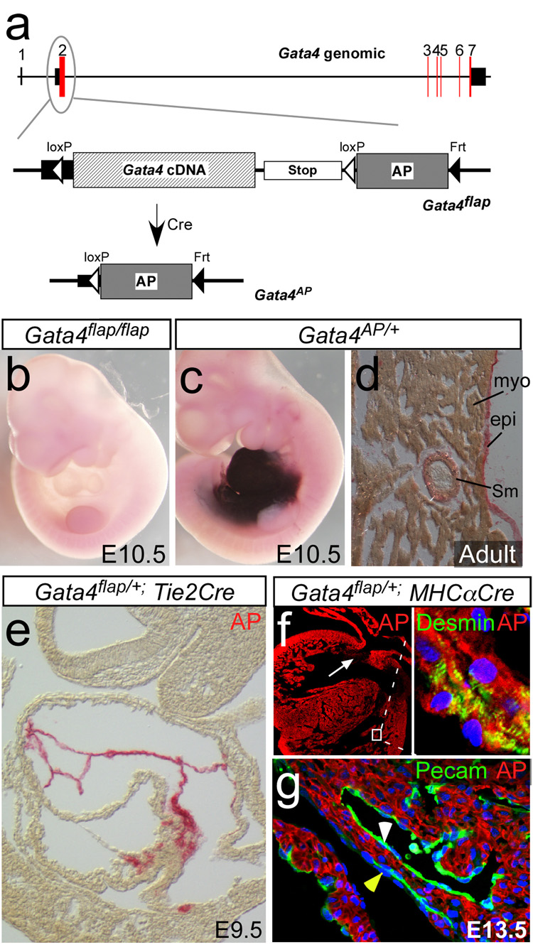
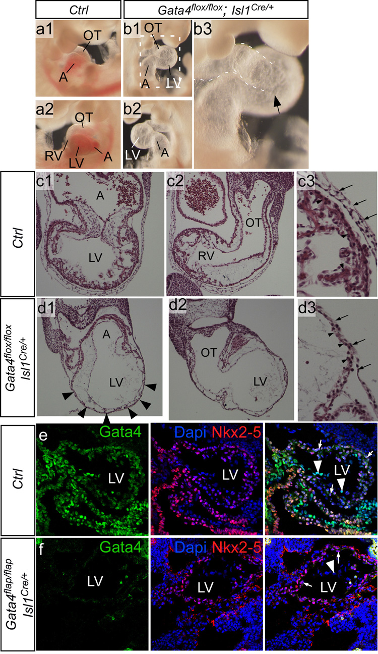
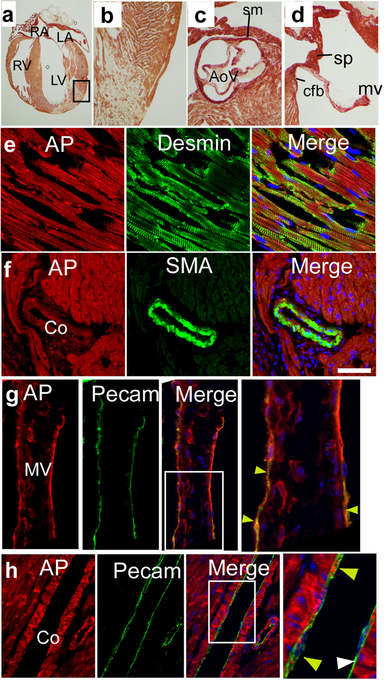
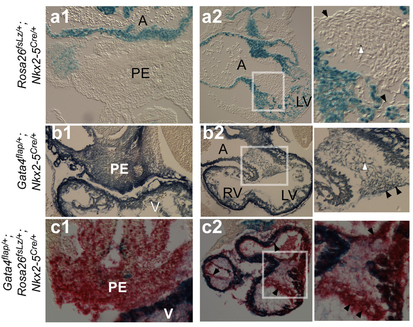
Isl1 and Nkx2-5-expressing cardiovascular progenitors play pivotal roles in cardiogenesis. Previously reported Cre-based fate-mapping studies showed that Isl1 progenitors contribute predominantly to the derivatives of the second heart field, and Nkx2-5 progenitors contributed mainly to the cardiomyocyte lineage. However, partial recombination of Cre reporter genes can complicate interpretation of Cre fate-mapping experiments. We found that a Gata4-based Cre-activated reporter was recombined by Isl1(Cre) and Nkx2-5(Cre) in a substantially broader domain than previously reported using standard Cre-activated reporters. The expanded Isl1 and Nkx2-5 cardiac fate maps were remarkably similar, and included extensive contributions to cardiomyocyte, endocardial, and smooth muscle lineages in all four cardiac chambers. These data indicate that Isl1 is expressed in progenitors of both primary and secondary heart fields, and that Nkx2-5 is expressed in progenitors of cardiac endothelium and smooth muscle, in addition to cardiomyocytes. These results have important implications for our understanding of cardiac lineage diversification in vivo, and for the interpretation of Cre-based fate maps.


Figures

Figure 1
Figure 1. Gata4flap, a reporter of cardiac Cre activity
a. Schematic depicting the structure of the Gata4 genomic locus, and the knockin Gata4flap allele. Red boxes indicate coding regions, and black boxes untranslated regions. AP, human placental alkaline phosphatase. b. Lack of AP activity in Gata4flap/flap embryos. c. AP activity in Gata4AP/+ embryo. d. AP expression in Gata4AP/+ adult heart. e. Tie2Cre activated Gata4flap in the endocardium but not myocardium. f–g. MHC〈Cre recombined Gata4flap selectively in cardiomyocytes (Myo). Cells of the endocardium (white arrowhead, g), epicardium (yellow arrowhead, g) and endocardial cushions (white arrow, f) were not recombined. AP was detected with BCIP/NBT in b,c and Permanent Red in d–g.
Figure 2
Figure 2. Developmental fates of Isl1+ progenitors in the developing heart
E9.5 Rosa26fsLz/+; Isl1Cre/+ (a), Gata4flap/+; Isl1Cre+ (b), and Gata4flap/+; Rosa26fsLz/+; Isl1Cre/+ (c) embryos were stained for β-galactosidase (blue, a), AP (red, b), or both (blue and red, c; overlap appears as dark blue). Embryos were then transversely sectioned. Note that Gata4flap was activated much more extensively than Rosa26fsLz in LV and AV cushion (askterisk and inset, a2 vs b2). Black arrowheads indicate endocardium marked by Isl1Cre. Arrows indicate LV myocardium, which strongly expressed AP but only expressed LacZ in a mosaic pattern. Abbreviations are as in Figure 1.
Figure 3
Figure 3. Inactivation of Gata4 by Isl1Cre
a–b. Gross morphology of control and mutant embryos at E10.5. Mutant embryos were growth retarded and often had large pericardial effusions (not shown). Removal of the pericardial sac showed a bulbous and translucent appearing LV, and absence or severe hypoplasia of the RV (arrow, b3). c–d. Histological transverse sections of E10.5 control and mutant embryos. The mutant embryos had severe thinning of the LV myocardium and absence of myocardial trabeculation (arrowheads, d1). The morphological RV was severely hypoplastic (not shown) to absent (d2). c3-d3 show higher magnification of LV myocardium. Arrows indicate pericardium, arrowheads LV myocardium. A, atrium. LV and RV, left and right ventricle. OT, outflow tract. e–f. Loss of Gata4 immunoreactivity in Isl1Cre-recombined mutant LV. Sagittal frozen sections of control and Gata4flap/flap; Isl1Cre/+ mutant E9.5 littermate hearts. Gata4 was expressed in cardiomyocytes (arrows) and endothelium (arrowheads) of control LV. In Gata4flap/flap; Isl1Cre/+ hearts, Gata4 immunoreactivity was not detected in either cardiomyocytes or endothelium of LV. Nkx2–5 expression was unchanged in the mutant, demonstrating preservation of antigen integrity in mutant as well as control samples.
Figure 4
Figure 4. Developmental fates of Isl1+ progenitors in the postnatal heart
Frozen sections of postnatal Gata4flap/+; Isl1Cre/+ hearts, stained for AP activity (red) and lineage markers. Isl1Cre activated Gata4flap in cardiomyocytes (a–b), in the base of the aorta and in the semilunar valves (c), and in derivatives of the AV endocardial cushions (d). AP activity colocalized with markers of cardiomyocytes (desmin, e), coronary SMCs (smooth muscle α-actin (SMA), f), and valve and coronary ECs (Pecam, yellow arrowheads, g and h). Coronary ECs were mosaic for AP staining (white arrowheads indicate Pecam+ cells that were AP negative). AoV, aortic valve. cfb, central fibrous body. co, coronary. mv, mitral valve. sm, smooth muscle. sp, atrial septum primum.
Figure 5
Figure 5. Developmental fates of Nkx2–5+ progenitors in the developing heart
Transverse paraffin sections of embryos (a–b, E10.0; c, E9.5) stained in whole mount for LacZ (blue) or AP (BCIP/NBT in b, Permanent Red in c). Boxed regions of AV endocardial cushions are enlarged in right panels. Nkx2–5Cre activated both reporters in cardiomyocytes (dark blue). Nkx2–5Cre activation of Gata4flap was markedly more efficient than Rosa26fsLz in endocardium (black arrowhead), endocardial cushion mesenchyme (white arrowhead), and proepicardium (PE).
Figure 6
Figure 6. Developmental fates of Nkx2–5+ progenitors in the postnatal heart
Gata4flap fate map of Nkx2–5 in adult heart. Frozen sections were stained for AP activity (red) and lineage markers. Nkx2–5Cre activated Gata4flap in cardiomyocytes (a–b), in the base of the aorta and in the semilunar valves (c), and in derivatives of the AV endocardial cushions (d). AP activity colocalized with markers of cardiomyocytes (e), coronary smooth muscle (f), valve endocardium (yellow arrowheads, g), and coronary ECs (yellow arrowheads, h). Endothelium was mosaic for AP staining (white arrowheads indicate Pecam+ cells that were AP negative). ad, adventitial tissue around aortic root, AoR, aortic root, tv, tricuspid valve. Other abbreviations as in Figure 4.

References

    1. Abu-Issa R, Waldo K, Kirby ML. Heart fields: one, two or more? Dev Biol. 2004;272:281–285. - PubMed
    1. Agah R, Frenkel PA, French BA, Michael LH, Overbeek PA, Schneider MD. Gene recombination in postmitotic cells. Targeted expression of Cre recombinase provokes cardiac-restricted, site-specific rearrangement in adult ventricular muscle in vivo. J Clin Invest. 1997;100:169–179. - PMC - PubMed
    1. Arceci RJ, King AA, Simon MC, Orkin SH, Wilson DB. Mouse GATA-4: a retinoic acid-inducible GATA-binding transcription factor expressed in endodermally derived tissues and heart. Mol Cell Biol. 1993;13:2235–2246. - PMC - PubMed
    1. Brade T, Gessert S, Kuhl M, Pandur P. The amphibian second heart field: Xenopus islet-1 is required for cardiovascular development. Dev Biol. 2007 - PubMed
    1. Bruneau BG, Black BL. The heart's Da Vinci code: a Renaissance at Keystone. Development. 2007;134:1631–1633. - PubMed

Publication types

MeSH terms

LinkOut - more resources