. 2008 Dec;23(12):2782-90.
doi: 10.1093/humrep/den298. Epub 2008 Sep 4.

Estrogen receptor related beta is expressed in human endometrium throughout the normal menstrual cycle

Affiliations

Estrogen receptor related beta is expressed in human endometrium throughout the normal menstrual cycle

Vincent Bombail et al. Hum Reprod. 2008 Dec.

Abstract

Background: Estrogen receptor related beta (ERRbeta, ESRRB/NR3B2) is an orphan receptor that shares significant sequence homology with estrogen receptors ERalpha and ERbeta. ERR family members are reported to exhibit constitutive transcriptional activity; however, little is known about the biological function of ERRbeta. In an attempt to delineate its role, we examined expression of ERRbeta in normal human endometrium, a tissue that undergoes cyclic remodelling under the influence of estrogen and progesterone.

Methods: Well-characterized endometrial tissue (n = 31), including full-thickness biopsies, was obtained from women with regular menstrual cycles. RT-PCR was used to measure mRNA encoding ERRbeta, the peroxisome proliferator activated receptor gamma coactivators (PGC)-1alpha and beta and to determine whether ERRbeta splice variant mRNAs were expressed. ERRbeta was immunolocalized using both single and double antibody immunohistochemistry.

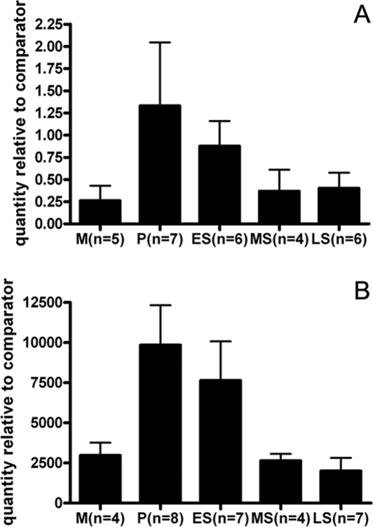
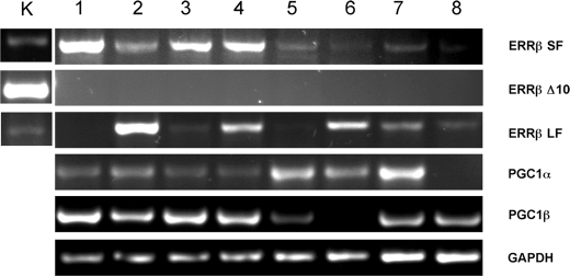
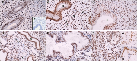
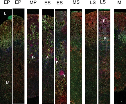
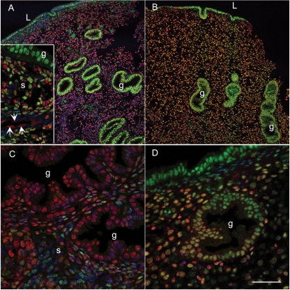
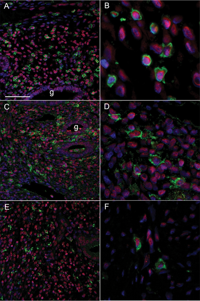
Results: Total ERRbeta mRNA appeared higher in proliferative phase samples but results did not reach significance. Transcripts corresponding to the long- and short-splice variants of ERRbeta as well as PGC1alpha and beta were detected but ERRbetaDelta10 was absent. ERRbeta protein was localized to cell nuclei within multiple endometrial cell types including the glands, stroma, endothelium and immune cells, including uterine natural killer (uNK) cells and macrophages. Fluorescent immunohistochemistry revealed that some cells co-expressed ERRbeta and ERalpha or ERbeta, for example, endothelial and uNK cells were ERRbeta+/ERbeta+.

Conclusions: ERRbeta mRNA and protein are expressed in healthy human endometrium. Further studies are warranted to characterize the functional impact of ERRbeta on endometrial biology.


Figures

Figure 1:
Figure 1:
Detection of ERRβ mRNAs in endometrial tissue using qRT–PCR. Expression of ERRβ (A) and ERα (B) mRNAs in human endometrial samples recovered during the normal cycle. RNA was extracted from pipelle biopsies taken from patients at different stages of the cycle, mRNA was evaluated using qRT–PCR. Data are expressed relative to an internal control and was compared using a one-way analysis of variance for ERα (P = 0.0052) or a Kruskal–Wallis test for ERRβ (P = 0.679). Data are mean ± SE. M, menstrual; P, proliferative; ES, early secretory; MS, mid-secretory; LS, late secretory.
Figure 2:
Figure 2:
Evidence that both long and short forms of ERRβ and the nuclear receptor coactivators PGC1α and PGC1β are present in normal endometrium. RT–PCR analysis of RNA from kidney (K), proliferative (lanes 1–4) and mid-secretory (lane 5–8) phase endometrium. The abbreviations on the right-hand side indentify DNA amplied with primers specific for the following: ERRβ short form (SF), ERRβΔ10 (Δ10), ERRβ long form (LF), PGC1α, PGC1β and GAPDH. The experiment was repeated three times and similar results were obtained on each occasion.
Figure 3:
Figure 3:
ERRβ protein is expressed in human endometrium throughout the cycle. Endometrial samples were dated as being from the following stages of the menstrual cycle; (A) Early proliferative, (B) late proliferative, (C) early secretory, (D) mid-secretory, (E) late secretory, (F) menstrual. (G and H) Immunopositive staining of cell nuclei in breast cancer and first trimester placenta, respectively (positive controls). The arrows point towards the endothelial cells of the spiral arterioles. The inset (A′) shows a section incubated with antibody pre-absorbed with the blocking peptide. Magnifications all ×20, bar in panel A′ is 50 microns and applies to all other images.
Figure 4:
Figure 4:
Full thickness endometrial biopsies taken throughout the menstrual cycle reveal differences in the expression of ERα and ERRβ proteins. ERRβ (red) was detected in cell nuclei in the functional layer (F) closest to the lumen (L) of the uterus. Tissues were dated as originating during the following phases of the cycle: EP, early proliferative; MP, mid-proliferative; ES, early secretory; MS, mid-secretory; LS, late secretory; M menstrual. Immunopositive staining for ERα (green) was particularly intense in cells lining the glands(g) during MP and ES phases. Note that groups of ERRβ positive cells (arrowheads) within the basal layer of the endometrium. The positions of the basal (B) and myometrial (M) layers are indicated.
Figure 5:
Figure 5:
Co-expression of ERα, ERβ and ERRβ proteins in functional layer of the endometrium during the normal menstrual cycle. Co-localization of ERRβ (red) with ERα (A and C) or ERβ (B and D) (both green); counterstained with 4’,6-diamidino-2-phenylindole (DAPI) (blue), yellow indicates colocalization. (A) Section from mid-proliferative endometrium with intense immunostaining for ERα in the glandular epithelium (g); inset shows high-power magnification to highlight mixed cell population in the stroma with cells including endothelial cells lining blood vessels (arrows) that express ERRβ but not ERα (red nuclei); (B) mid-secretory endometrium (same sample as in A) with prominent expression of ERβ1 in glandular epithelium and cells lining the lumen (L); (C) section from late secretory endometrium (code 2232) with reduced expression of ERα revealing expression of ERRβ as red nuclei; (D) ERRβ and ERβ in late secretory endometrium (2232), note overlapping pattern of expression (yellow nuclei). Panels A and B ×10 and C and D ×40, scale bar = 50 microns and applies to panels C and D.
Figure 6:
Figure 6:
ERRβ protein is expressed in immune cell populations within the human endometrium. Co-localization of ERRβ (red nuclei) with surface markers for selected immune cell populations (all green), counterstain for cell nuclei was DAPI (blue). (A and B) CD45 leukocyte common antigen (green); (C and D) CD56 (green), uNK cells; (E and F) CD68 (green), uterine macrophages. Magnification panels A, C and E all ×20, scale bar 100 microns; panels B, D and F are cropped high-power views from the same samples.

References

    1. Aghajanova L, Hamilton AE, Giudice LC. Uterine receptivity to human embryonic implantation: histology, biomarkers, and transcriptomics. Semin Cell Dev Biol. 2008;19:204–211. - PMC - PubMed
    1. Aranda A, Pascual A. Nuclear hormone receptors and gene expression. Physiol Rev. 2001;81:1269–1304. - PubMed
    1. Barry JB, Giguere V. Epidermal growth factor-induced signaling in breast cancer cells results in selective target gene activation by orphan nuclear receptor estrogen-related receptor alpha. Cancer Res. 2005;65:6120–6129. - PubMed
    1. Critchley HOD, Brenner RM, Henderson TA, Williams K, Nayak NR, Slayden OD, Millar MR, Saunders PTK. Estrogen receptor beta, but not estrogen receptor alpha, is present in the vascular endothelium of the human and nonhuman primate endometrium. J Clin Endocrinol Metab. 2001;86:1370–1378. - PubMed
    1. Critchley HO, Henderson TA, Kelly RW, Scobie GS, Evans LR, Groome NP, Saunders PT. Wild-type estrogen receptor (ERbeta1) and the splice variant (ERbetacx/beta2) are both expressed within the human endometrium throughout the normal menstrual cycle. J Clin Endocrinol Metab. 2002;87:5265–5273. - PubMed

Publication types

MeSH terms