. 2008 Nov 21;283(47):32527-33.
doi: 10.1074/jbc.M803059200. Epub 2008 Sep 5.

Endoglin promotes transforming growth factor beta-mediated Smad 1/5/8 signaling and inhibits endothelial cell migration through its association with GIPC

Affiliations

Endoglin promotes transforming growth factor beta-mediated Smad 1/5/8 signaling and inhibits endothelial cell migration through its association with GIPC

Nam Y Lee et al. J Biol Chem. .

Abstract

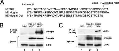
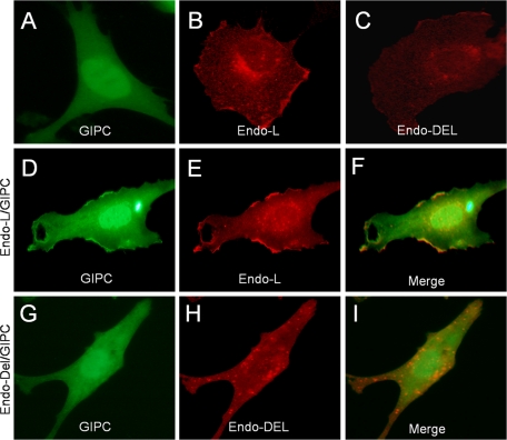
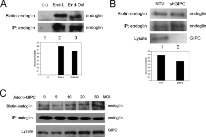
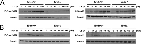
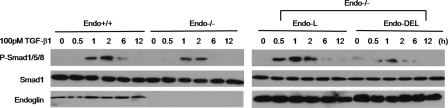
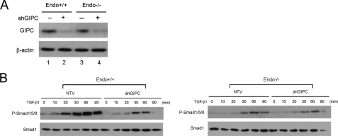
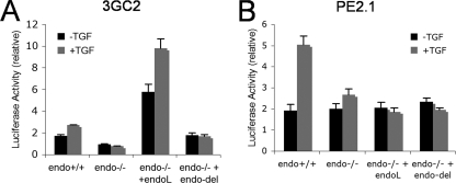
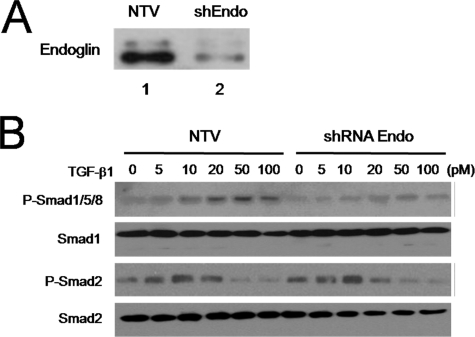
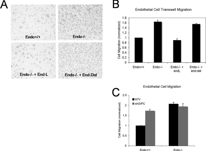
Transforming growth factor beta (TGF-beta) signals through two distinct pathways to regulate endothelial cell proliferation, migration, and angiogenesis, the ALK-1/Smad 1/5/8 and ALK-5/Smad2/3 pathways. Endoglin is a co-receptor predominantly expressed in endothelial cells that participates in TGFbeta-mediated signaling with ALK-1 and ALK-5 and regulates critical aspects of cellular and biological responses. The embryonic lethal phenotype of knock-out mice because of defects in angiogenesis and disease-causing mutations resulting in human vascular diseases both support essential roles for endoglin, ALK-1, and ALK-5 in the vasculature. However, the mechanism by which endoglin mediates TGF-beta signaling through ALK-1 and ALK-5 has remained elusive. Here we describe a novel interaction between endoglin and GIPC, a scaffolding protein known to regulate cell surface receptor expression and trafficking. Co-immunoprecipitation and immunofluorescence confocal studies both demonstrate a specific interaction between endoglin and GIPC in endothelial cells, mediated by a class I PDZ binding motif in the cytoplasmic domain of endoglin. Subcellular distribution studies demonstrate that endoglin recruits GIPC to the plasma membrane and co-localizes with GIPC in a TGFbeta-independent manner, with GIPC-promoting cell surface retention of endoglin. Endoglin specifically enhanced TGF-beta1-induced phosphorylation of Smad 1/5/8, increased a Smad 1/5/8 responsive promoter, and inhibited endothelial cell migration in a manner dependent on the ability of endoglin to interact with GIPC. These studies define a novel mechanism for the regulation of endoglin signaling and function in endothelial cells and demonstrate a new role for GIPC in TGF-beta signaling.


Figures

FIGURE 1.
FIGURE 1.
Endoglin interacts with GIPC. A, schematic representation of the cytoplasmic domains of human TβRIII, endoglin wild type, and endoglin-Del. B, anti-Flag immunoprecipitates were prepared from endo-/- MEECs expressing Flag-tagged GIPC with empty vector (lane 1), endoglin-Del (lane 2), and endoglin-L (lane 3). Upper panel, immunoblot of HA-tagged endoglin constructs co-precipitated with Flag-tagged GIPC. Middle and lower panels, immunoblots of endoglin and GIPC in total cell lysates, respectively. C, anti-HA immunoprecipitates were prepared from endo-/- MEECs expressing Flag-tagged GIPC with empty vector (lane 1), endoglin-Del (lane 2), and endoglin-L (lane 3). Upper panel, immunoblot of Flag-tagged GIPC co-precipitated with HA-tagged endoglin constructs. Middle and lower panels, immunoblots of endoglin and GIPC in total cell lysates, respectively. Data are representative of three independent experiments.
FIGURE 2.
FIGURE 2.
Recruitment and co-localization of GIPC with endoglin. Endo-/- MEECs transiently expressing GIPC-GFP alone (A), HA-tagged endoglin-L alone (B), HA-tagged endoglin-Del alone (C), GIPC-GFP with endoglin-L (D–F) or GIPC-GFP with endoglin-Del (G–I) were fixed, permeabilized, and immunostained for endoglin with endoglin antibody (P3D1) to visualize endoglin and GIPC localization using confocal imaging (60×). GIPC-GFP co-localizes with endoglin-L at distinct parts at or near the plasma membrane (D, E, and merged image in F), but not with endoglin-Del (G, H, and merged image I). Data are representative of three independent experiments.
FIGURE 3.
FIGURE 3.
GIPC stabilizes endoglin on the cell surface. A, an empty vector control (lane 1), endoglin-L (lane 2), or endoglin-Del (lane 3) were expressed in Endo-/- MEECs for 40 h prior to cell surface biotinylation. Cells were subsequently immunoprecipitated with P3D1 endoglin antibody, and then resolved on SDS-PAGE before being observed with streptavidin-conjugated HRP (upper panel, biotinylated cell surface endoglin, lower panel, immunoprecipitated total cellular endoglin). Cell surface biotinylated endoglin and endoglin-DEL levels were quantified by densitometry scan, normalized to total endoglin expression and represented in arbitrary units (lower panel). B, non-targeting vector (lane 1) or shRNA against GIPC (lane 2) were nucleofected into endo+/+ MEECs for 48 h prior to cell surface biotinylation followed by immunoprecipitation of endogenous endoglin (upper panel, biotinylated cell surface endoglin; middle panel, immunoprecipitated endogenous total cellular endoglin; lower panel, endogenous GIPC in cell lysate). Cell surface biotinylated endoglin levels were quantified by densitometry scan, normalized to total endoglin expression, and represented in arbitrary units (lower panel). C, after infection with adenovirus encoding GIPC at the indicated MOIs, HMEC-1 cells were cell surface biotinylated. Cells were subsequently immunoprecipitated with P3D1 endoglin antibody, then resolved on SDS-PAGE before being observed with streptavidin-conjugated HRP (upper panel, biotinylated cell surface endoglin; middle panel, immunoprecipitated total cellular endoglin). Expression levels of GIPC were confirmed in total cell lysate (lower panel). Data are representative of two to three independent experiments.
FIGURE 4.
FIGURE 4.
Endoglin promotes Smad1/5/8 activation. A, Endo+/+ and endo-/- MEECs were serum-starved for 6 h prior to treatment with TGF-β1 for 20 min at the indicated doses. Cells were harvested, the lysates normalized for protein levels via Bradford assay, and levels of Smad 1/5/8 and Smad 2 phosphorylation were assessed with phosphospecific Smad 1/5/8 and Smad 2 antibodies. Upper panels, immunoblot of phospho-Smad1/5/8 (left) and phospho-Smad 2 (right) levels. Lower panels, immunoblot of total Smad 1 (left) and Smad 2(right) levels. B, Endo+/+ and endo-/- MEECs were treated with 50 pm TGF-β1 for the indicated times and analyzed for Smad 1/5/8 and Smad 2 phosphorylation (upper panels, left and right, respectively). Lower panels, immunoblots of total Smad 1 (left) and Smad 2 (right) levels. Data are representative of three independent experiments.
FIGURE 5.
FIGURE 5.
Endoglin interacts with GIPC to promote Smad 1/5/8 activation. Endo+/+, endo-/- MEECs or endo-/- MEECs nucleofected with endoglin-L and endoglin-Del were serum-starved for 6 h prior to 100 pm TGF-β1 stimulation for the indicated times (in hours (h)). Cells were harvested and normalized for protein levels via Bradford, and levels of Smad 1/5/8 were assessed by use of phosphospecific Smad 1/5/8 antibodies. Upper panels, immunoblot of phospho-Smad1/5/8 levels. Middle panels, immunoblot of total Smad 1 levels. Lower panels, immunoblot of endoglin expression. Data are representative of three independent experiments.
FIGURE 6.
FIGURE 6.
GIPC knockdown attenuates Smad 1/5/8 activation. A, both endo+/+ and endo-/- MEECs were nucleofected with either non-targeting vector control (lanes 1 and 3) or shRNA against GIPC (lanes 2 and 4), and the expression of GIPC assessed by Western blot (upper panel) with β-actin assessed as a loading control (lower panel). B, 48 h post-nucleofection with either the non-targeting vector (NTV) or GIPC shRNA, cells were serum-starved for 6 h prior to 100 pm TGF-β1 treatment for the indicated time (hours (h)). Upper panels, immunoblot of phospho-Smad1/5/8 levels. Lower panels, immunoblot of total Smad 1 levels. Data are representative of two independent experiments.
FIGURE 7.
FIGURE 7.
Endoglin up-regulates Smad1 reporter gene activation. Endo+/+ and endo-/- MEECs were nucleofected with either (A) 3GC2 (a Smad 1 reporter) or (B) PE2.1 (a Smad 2 reporter) along with an empty vector, endoglin-L, or endoglin-Del construct. SV40 plasmid nucleofection was included in all the samples for Renilla activity (SV40) and used to control for transfection efficiency. The indicated error bars represent the S.E. from triplicates for each of the represented experimental conditions. Data shown are representative of three independent experiments.
FIGURE 8.
FIGURE 8.
Endoglin promotes Smad 1/5/8 activation in HMEC-1 cells. A, endoglin expression was assessed in HMEC-1s 72 h after infection with an adenovirus containing a non-targeting vector (NTV, lane 1) or shEndoglin (lane 2). B, phosphorylation levels of Smad 1/5/8 and Smad 2 were assessed via phosphospecific Smad 1/5/8 and Smad 2 antibodies 20 min after TGF-β1 treatment. The first and third panels indicate the phosphorylation of Smad 1/5/8 and Smad 2, respectively. The second and fourth panels indicate the total Smad 1 and Smad 2, respectively. All cells were harvested and normalized for protein levels via Bradford. Data shown are representative of three independent experiments.
FIGURE 9.
FIGURE 9.
Endoglin interacts with GIPC to regulate migration. Endo+/+ and endo-/- MEECs were plated on transwells coated with 0.02% gelatin and assessed for migration through the transwells 12 h later. A, migrated cells found on the bottom side of the membrane were fixed, stained for their nuclei, and imaged for endo+/+ (upper left panel), endo-/- (upper right), endo-/- with endoglin-L expression (lower left), and endo-/- with endoglin-Del (lower right). B, number of migrated cells on the membrane bottom (shown as stained nuclei) were counted using Image J software. Cell migration is represented as a percentage, normalized to endo+/+ MEECs, from triplicates for each of the four independent experiments. The error bars indicate the standard error of the mean of the percentage of the migrated cells. C, transwell migration analyses upon shRNA-mediated GIPC knockdown. 48 h post-nucleofection with either non-targeting vector (NTV) or shRNA (shGIPC) endo+/+ and endo-/- MEECs were plated onto transwells for 12 h. Cells were fixed, stained, and quantitated as above.

References

    1. Massague, J. (1998) Annu. Rev. Biochem. 67, 753-791 - PubMed
    1. Cheifetz, S., Andres, J. L. and Massague, J. (1988) J. Biol. Chem. 263 16984-16991 - PubMed
    1. Cheifetz, S., Bellón, T., Calés, C., Vera, S., Bernabeu, C., Massagué, J., and Letarte, M. (1992) J. Biol. Chem. 267 19027-19030 - PubMed
    1. Barbara, N. P., Wrana, J. L., and Letarte, M. (1999) J. Biol. Chem. 274 584-594 - PubMed
    1. Ten Dijke, P., Goumans, M. J., Itoh, F., and Itoh, S. (2002) J. Cell. Physiol. 191 1-16 - PubMed

Publication types

MeSH terms

Substances

LinkOut - more resources