. 2008 Nov;190(22):7441-52.
doi: 10.1128/JB.00584-08. Epub 2008 Sep 5.

The bile response repressor BreR regulates expression of the Vibrio cholerae breAB efflux system operon

Affiliations

The bile response repressor BreR regulates expression of the Vibrio cholerae breAB efflux system operon

Francisca A Cerda-Maira et al. J Bacteriol. 2008 Nov.

Abstract

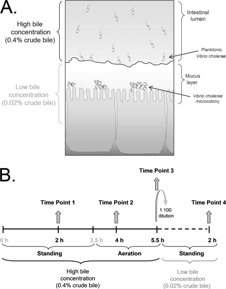
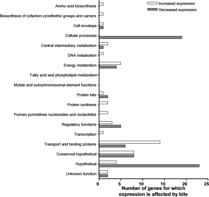
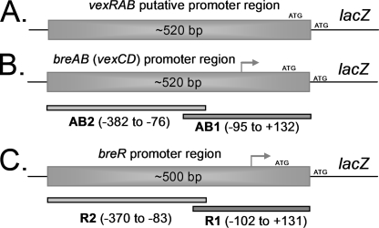
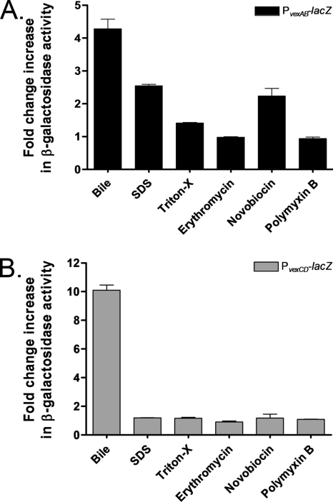
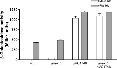
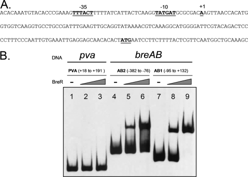
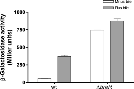
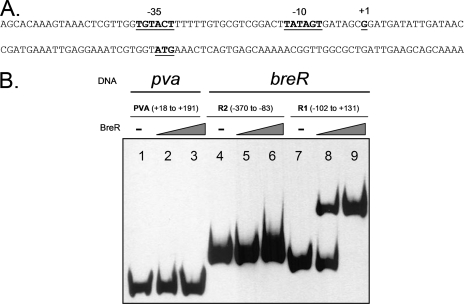
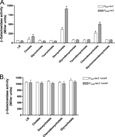
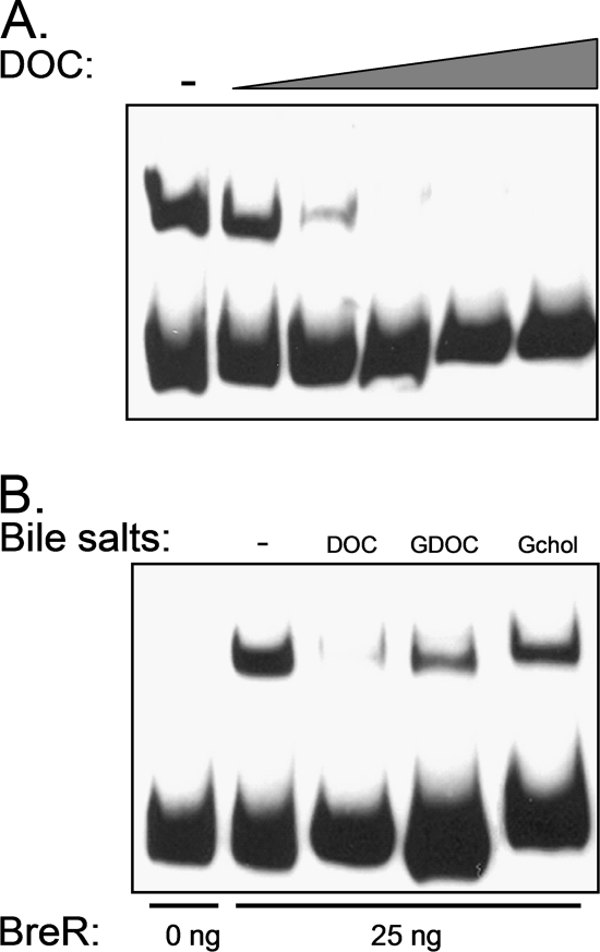
Enteric pathogens have developed several resistance mechanisms to survive the antimicrobial action of bile. We investigated the transcriptional profile of Vibrio cholerae O1 El Tor strain C6706 under virulence gene-inducing conditions in the presence and absence of bile. Microarray analysis revealed that the expression of 119 genes was affected by bile. The mRNA levels of genes encoding proteins involved in transport were increased in the presence of bile, whereas the mRNA levels of genes encoding proteins involved in pathogenesis and chemotaxis were decreased. This study identified genes encoding transcriptional regulators from the TetR family (vexR and breR) and multidrug efflux pumps from the resistance-nodulation-cell division superfamily (vexB and vexD [herein renamed breB]) that were induced in response to bile. Further analysis regarding vexAB and breAB expression in the presence of various antimicrobial compounds established that vexAB was induced in the presence of bile, sodium dodecyl sulfate, or novobiocin and that the induction of breAB was specific to bile. BreR is a direct repressor of the breAB promoter and is able to regulate its own expression, as demonstrated by transcriptional and electrophoretic mobility shift assays (EMSA). The expression of breR and breAB is induced in the presence of the bile salts cholate, deoxycholate, and chenodeoxycholate, and EMSA showed that deoxycholate is able to abolish the formation of BreR-P(breR) complexes. We propose that deoxycholate is able to interact with BreR and induce a conformational change that interferes with the DNA binding ability of BreR, resulting in breAB and breR expression. These results provide new insight into a transcriptional regulator and a transport system that likely play essential roles in the ability of V. cholerae to resist the action of bile in the host.


Figures

FIG. 1.
FIG. 1.
(A) Model illustrating the effect of bile on V. cholerae colonization based on a hypothesis proposed by Schuhmacher and Klose (48). When V. cholerae is within the lumen of the intestine, the high bile concentration inhibits the transcription of the virulence genes and induces motility and/or chemotaxis to mobilize the bacterium into the mucus layer. Upon migration through the mucus layer, where the bile concentration is low, motility and/or chemotaxis is inhibited and virulence gene expression is induced, facilitating the colonization of the epithelial cells by V. cholerae. (B) Schematic representation of the microarray experimental design.
FIG. 2.
FIG. 2.
Distribution of V. cholerae El Tor genes differentially expressed in response to crude bile as classified within clusters of orthologous groups assigned by the TIGR database.
FIG. 3.
FIG. 3.
Diagrams showing the promoter regions and fragments employed to generate lacZ transcriptional fusions and DIG-dUTP-labeled fragments. (A) Fragment of ∼520 bp from the upstream region of the putative vexAB ATG start codon. (B) breAB (vexCD) promoter region. Fragments AB1 and AB2 were used for EMSA. (C) breR promoter region. The R1 and R2 fragments were used for EMSA. The breAB and breR transcriptional start sites are indicated by gray arrows.
FIG. 4.
FIG. 4.
Induction of PvexRAB-lacZ (A) and PvexCD-lacZ (PbreAB-lacZ) (B) expression by different compounds. β-Galactosidase expression was measured by growing the strains in the absence or presence of subinhibitory concentrations of crude bile (0.4%), SDS (300 μM), Triton X-100 (150 μg/ml), erythromycin (0.1 μM), novobiocin (0.1 μM), or polymyxin B (5 U/ml) in LB at 37°C until the OD600 of the cultures reached 0.8 to 1.0. The amount of change (n-fold) in β-galactosidase activity was calculated by dividing the level of β-galactosidase activity obtained in the presence of each compound by the activity obtained in the absence of the compound. The results shown are from three independent experiments. Error bars indicate standard deviations.
FIG. 5.
FIG. 5.
Induction of PbreAB-lacZ expression in various strain backgrounds by crude bile. β-Galactosidase expression was measured by growing the strains in the absence or presence of 0.4% crude bile in LB at 37°C until the OD600 of the cultures reached 0.8 to 1.0. The results shown are from three independent experiments. Error bars indicate standard deviations.
FIG. 6.
FIG. 6.
Specific binding of BreR to the breAB promoter region. (A) Nucleotide sequence of the breAB promoter. The position of the transcriptional start site for breAB was determined by 5′ RACE. The transcriptional start site (+1), ATG start codon, and putative −35 and −10 regions are boldfaced and underlined. (B) EMSA was performed with the control DNA fragment from the pva promoter (25) (lanes 1 to 3) or the breAB promoter fragments AB1 and AB2 (lanes 4 to 9). DIG-dUTP-labeled DNA (10 ng) was incubated with 0 ng (lanes 1, 4, and 7), 50 ng (lanes 2, 5, and 8), or 250 ng (lanes 3, 6, and 9) of BreR-His6 prior to electrophoresis. −, no BreR.
FIG. 7.
FIG. 7.
Induction of PbreR-lacZ expression by crude bile and breR autoregulation. β-Galactosidase activity was measured by growing the strains in the absence or presence of 0.4% crude bile in LB at 37°C until the OD600 of the cultures reached 0.8 to 1.0. The results shown are from three independent experiments. Error bars indicate standard deviations. wt, wild type.
FIG. 8.
FIG. 8.
BreR interaction with the breR promoter region. (A) The breR promoter nucleotide sequence is shown. 5′ RACE was utilized to determine the position of the transcriptional start site for breR. The transcriptional start site (+1), ATG start codon, and putative −35 and −10 regions are boldfaced and underlined. (B) EMSA was performed with the control DNA fragment from the pva promoter (25) (lanes 1 to 3) or the breR promoter fragments R1 and R2 (lanes 4 to 9). In the DNA binding assay, the DIG-dUTP-labeled DNA (10 ng) was incubated with 0 ng (lanes 1, 4, and 7), 50 ng (lanes 2, 5, and 8), or 250 ng (lanes 3, 6, and 9) of BreR-His6.
FIG. 9.
FIG. 9.
Influence of different bile salts on the expression of PbreAB-lacZ and PbreR-lacZ as determined by β-galactosidase assays. Strains carrying the PbreR-lacZ or the PbreAB-lacZ fusion were grown in LB in the absence or presence of a subinhibitory concentration (300 μM) of different bile salts at 37°C until the OD600 of the cultures reached 0.8 to 1.0. The results shown are from three independent experiments. Error bars indicate standard deviations. Asterisks indicate statistically significant differences from the LB control. (A) Induction of PbreAB-lacZ and PbreR-lacZ fusions in the presence of different bile salts. (B) Expression of PbreAB-lacZ and PbreR-lacZ in a ΔbreR background in response to different bile salts.
FIG. 10.
FIG. 10.
Effect of deoxycholate on the DNA binding activity of BreR. (A) Titration of deoxycholate (DOC) to determine the concentration that prevents the formation of BreR-R1 complexes. EMSA was performed with the R1 fragment and 25 ng of BreR-His6. DIG-dUTP-labeled DNA (10 ng) was incubated with increasing concentrations (0, 5, 10, 20, 40, and 80 mM) of DOC prior to electrophoresis. −, no DOC. (B) EMSA analysis showing the disruption of the BreR-DNA complex in the presence of DOC but not glycodeoxycholate (GDOC) or glycocholate (Gchol). EMSA was performed with the R1 fragment and 0 or 25 ng of BreR-His6 (lanes − to Gchol). DIG-dUTP-labeled DNA (10 ng) was incubated with no bile salts (−), 10 mM DOC, 10 mM GDOC, or 10 mM Gchol.

References

    1. Aires, J. R., T. Kohler, H. Nikaido, and P. Plesiat. 1999. Involvement of an active efflux system in the natural resistance of Pseudomonas aeruginosa to aminoglycosides. Antimicrob. Agents Chemother. 432624-2628. - PMC - PubMed
    1. Begley, M., C. G. Gahan, and C. Hill. 2005. The interaction between bacteria and bile. FEMS Microbiol. Rev. 29625-651. - PubMed
    1. Bina, J. E., and J. J. Mekalanos. 2001. Vibrio cholerae tolC is required for bile resistance and colonization. Infect. Immun. 694681-4685. - PMC - PubMed
    1. Bina, J. E., D. Provenzano, C. Wang, X. R. Bina, and J. J. Mekalanos. 2006. Characterization of the Vibrio cholerae vexAB and vexCD efflux systems. Arch. Microbiol. 186171-181. - PubMed
    1. Blanco, C., M. Mata-Gilsinger, and P. Ritzenthaler. 1985. The use of gene fusions to study the expression of uidR, a negative regulatory gene of Escherichia coli K-12. Gene 36159-167. - PubMed

Publication types

MeSH terms