. 2008 Oct;74(20):6427-36.
doi: 10.1128/AEM.00815-08. Epub 2008 Sep 5.

In situ detection of freshwater fungi in an alpine stream by new taxon-specific fluorescence in situ hybridization probes

Affiliations

In situ detection of freshwater fungi in an alpine stream by new taxon-specific fluorescence in situ hybridization probes

Christiane Baschien et al. Appl Environ Microbiol. 2008 Oct.

Abstract

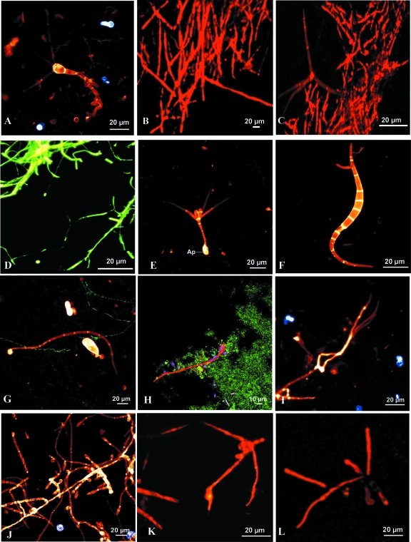
New rRNA-targeting oligonucleotide probes permitted the fluorescence in situ hybridization (FISH) identification of freshwater fungi in an Austrian second-order alpine stream. Based on computer-assisted comparative sequence analysis, nine taxon-specific probes were designed and evaluated by whole-fungus hybridizations. Oligonucleotide probe MY1574, specific for a wide range of Eumycota, and the genus (Tetracladium)-specific probe TCLAD1395, as well as the species-specific probes ALacumi1698 (Alatospora acuminata), TRIang322 (Tricladium angulatum), and Alongi340 (Anguillospora longissima), are targeted against 18S rRNA, whereas probes TmarchB10, TmarchC1_1, TmarchC1_2, and AlongiB16 are targeted against the 28S rRNA of Tetracladium marchalianum and Anguillospora longissima, respectively. After 2 weeks and 3 months of exposure of polyethylene slides in the stream, attached germinating conidia and growing hyphae of freshwater fungi were accessible for FISH. Growing hyphae and germinating conidia on leaves and in membrane cages were also visualized by the new FISH probes.


Figures

FIG. 1.
FIG. 1.
FISH with new specific oligonucleotide probes. (A) CLSM photomicrograph of a conidium and conidiophore of Clavariopsis aquatica growing out of a Fagus sylvatica leaf collected from the Oberer Seebach, shown after hybridization with Cy3-labeled probe MY1574. (B) CLSM photomicrograph showing FISH of Tetracladium marchalianum with specific 28S rRNA-targeting Cy3-labeled probe TmarchC1_1. (C) CLSM photomicrograph of Alatospora acuminata hybridized with 18S rRNA-targeting Cy3-labeled probe ALacumi1698. (D) Epifluorescence photomicrograph of Anguillospora longissima after FISH with 18S rRNA-targeting Cy3-labeled probe Alongi340. (E) Conidium of Tetracladium marchalianum attached to a PE slide exposed for 2 weeks within the Oberer Seebach (Lunz; autumn 2000) and then hybridized with Cy3-labeled probe MY1574 and simultaneously stained with calcofluor white; Ap, appressorium. (F) Germinating conidium of Anguillospora cf. crassa (similar to that of A. crassa or to the broad conidium of A. longissima) attached to a PE slide (after a 2-week exposure in autumn 2000), shown after FISH with Cy3-labeled probe MY1574 and simultaneous staining with calcofluor white. (G) Conidium of Anguillospora cf. filiformis attached to a PE slide exposed for 2 weeks within the Oberer Seebach (Lunz; autumn 2000) and then hybridized with Cy3-labeled probe MY1574 and simultaneously stained with calcofluor white, along with bacteria hybridized with Oregon green-labeled probe EUB338. (H) Biofilm obtained on a PE slide exposed for 3 months (September to November 2000) within the Oberer Seebach and analyzed with Oregon green-labeled probe EUB338 (green) and Cy3-labeled probe MY1574 (red). Also visible are autofluorescing diatoms. (I) CLSM photomicrograph of an unidentified fungus producing filiform phialoconidia on a leaf of Fagus sylvatica exposed for 2 weeks in the Oberer Seebach (autumn 2000), shown after hybridization with Cy3-labeled probe MY1574. (J) CLSM photomicrograph of fungal hyphae and phialids on a Fagus sylvatica leaf exposed for 2 weeks in the Oberer Seebach (autumn 2000), shown after hybridization with Cy3-labeled probe MY1574 and simultaneous staining with calcofluor white. (K) Germinating conidium of Tetracladium marchalianum after 2 days of exposure, probed with TmarchB10. (L) Germinating conidium of Varicosporium elodeae after 2 days of exposure, probed with MY1574.

Similar articles

Cited by

References

    1. Amann, R. I., W. Ludwig, and K.-H. Schleifer. 1995. Phylogenetic identification and in situ detection of individual microbial cells without cultivation. Microb. Rev. 59:143-169. - PMC - PubMed
    1. Arsuffi, T. I., and K. Suberkropp. 1989. Selective feeding by shredders on leaf-colonizing stream fungi: comparison on macroinvertebrate taxa. Oecologia 79:30-37. - PubMed
    1. Baker, B. J., M. A. Lutz, S. C. Dawson, P. L. Bond, and J. F. Banfield. 2004. Metabolically active eukaryotic communities in extremely acidic mine drainage. Appl. Environ. Microbiol. 70:6264-6271. - PMC - PubMed
    1. Bärlocher, F. 1982. Conidium production from leaves and needles in four streams. Can. J. Bot. 60:1487-1494.
    1. Bärlocher, F. 1992. Community organisation, p. 38-69. In F. Bärlocher (ed.), The ecology of aquatic hyphomycetes. Springer Verlag, Berlin, Germany.

Publication types

Associated data

LinkOut - more resources