. 2008 Nov;19(11):2150-8.
doi: 10.1681/ASN.2008020233. Epub 2008 Sep 5.

Podocyte-specific deletion of dicer alters cytoskeletal dynamics and causes glomerular disease

Affiliations

Podocyte-specific deletion of dicer alters cytoskeletal dynamics and causes glomerular disease

Scott J Harvey et al. J Am Soc Nephrol. 2008 Nov.

Abstract

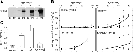
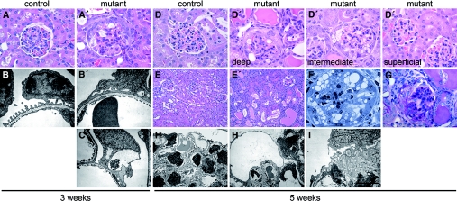
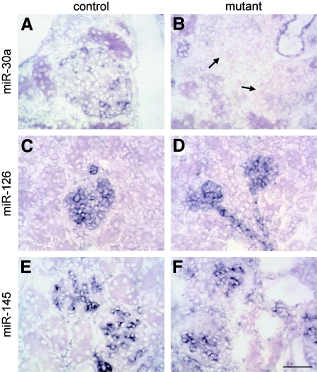
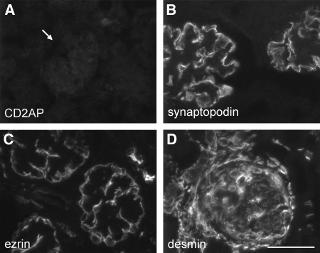
MicroRNAs (miRNAs) regulate gene expression by binding the 3' untranslated region of mRNAs. To define their role in glomerular function, miRNA biogenesis was disrupted in mouse podocytes using a conditional Dicer allele. Mutant mice developed proteinuria by 3 wk after birth and progressed rapidly to end-stage kidney disease. Podocyte pathology included effacement, vacuolization, and hypertrophy with crescent formation. Despite normal expression of WT1, podocytes underwent dedifferentiation, exemplified by cytoskeletal disruption with early transcriptional downregulation of synaptopodin. These abnormalities differed from Cd2ap(-/-) mice, indicating they were not a general consequence of glomerular disease. Glomerular labeling of ezrin, moesin, and gelsolin was altered at 3 wk, but expression of nestin and alpha-actinin was unchanged. Abnormal cell proliferation or apoptosis was not responsible for the glomerular injury. Mutant podocytes were incapable of synthesizing mature miRNA, as revealed by their loss of miR-30a. In contrast, expression of glomerular endothelial and mesangial cell miRNAs (miR-126 and miR-145, respectively) was unchanged. These findings demonstrate a critical role for miRNA in glomerular function and suggest a pathway that may participate in the pathogenesis of kidney diseases of podocyte origin. The unique architecture of podocytes may make them especially susceptible to cytoskeletal alterations initiated by aberrant miRNA dynamics.


Figures

Figure 1.
Figure 1.
Podocyte-specific Dicer knockout mice develop proteinuria by 3 wk and progress rapidly to end-stage kidney disease. (A) Analysis of urine from controls (C) and Dicerfl/fl;2.5P-Cre mice (fl/fl) by SDS-PAGE reveals progressive albuminuria in mutants by approximately 3 wk. (B) Dicerfl/fl;2.5P-Cre mutants show a corresponding increase in urinary protein/creatinine ratios and progress to end-stage kidney disease by approximately 6 wk. The phenotype was more severe in DicerΔ/fl;2.5P-Cre mutants (Δ/fl) carrying a null allele, and by 5 wk, most showed signs of uremia, including ascites, weight loss, and lethargy uncommon in Dicerfl/fl;2.5P-Cre mice at this age. Conversely, the phenotype was less severe in Dicerfl/fl;2.5P-Cre; R26R mutants (fl/fl; R26R) carrying a Cre-dependent reporter. The data include serial measurements on the number of mice indicated. (C) Blood urea nitrogen values were comparable in Dicerfl/fl;2.5P-Cre;R26R mutants and controls at 5 to 6 wk, whereas the differences between all other genotypes were statistically significant (P < 0.05). Disease progression is correlated with the number of “floxed” genomic loci, likely reflecting the limited efficiency of conditional targeting. Germline and podocyte-specific Dicer heterozygotes (DicerΔ/+ and Dicerfl/+;2.5P-Cre, respectively) had normal urinary protein/creatinine ratios and no overt phenotype.
Figure 2.
Figure 2.
Dicer is essential for maintenance of podocyte structure. (A and A′) Mutants at 3 wk show focal hypertrophy and vacuolization of epithelial cells in corticomedullary glomeruli, as well as pseudocrescents. (B, B′, and C′) Electron microscopy (EM) reveals segmental podocyte foot process effacement and vacuolization in affected glomeruli. (D and D′) The pathology is zonal in nature, and even at 5 wk, deep and intermediate glomeruli show changes, whereas superficial glomeruli are normal. (E′, F, and G) At advanced stages, mutants have tubular dilation with luminal protein casts and nuclei (E′), prominent vacuolization of glomerular epithelial cells (F), and FSGS and periglomerular fibrosis (G). (H, H′, and I) EM at 5 wk reveals global podocyte foot process effacement and prominent vacuolization (H′), whereas some glomeruli show collapsing lesions (I). Bar = 50 μm in A, A′, D, and D′; 150 μm in E and E′; 30 μm in F; 1.45 μm in B and B′; 3.13 μm in C and I; and 7.41 μm in H and H′; n ≥ 13 and n ≥ 4 mice for each light microscopy and EM panel, respectively.
Figure 3.
Figure 3.
miRNA processing is abolished in mutant podocytes. miRNA expression was evaluated in the kidneys of 3-wk-old by in situ hybridization. (A) miR-30a was expressed by collecting duct epithelium and podocytes in controls. (B) Podocytes in mutant glomeruli (arrows) were negative, a finding consistent with loss of Dicer activity, whereas tubular labeling was comparable to controls. This involved all podocytes, including those populating superficial glomeruli that show no pathology. (C and D) Signals for miR-126 were detected in glomerular and peritubular capillary endothelial cells in controls (C), and labeling was comparable in mutants (D). (E and F) miR-145 was expressed by mesangial and vascular smooth muscle cells in normal kidney (E), and labeling was indistinguishable in mutants (F). Bar = 50 μm; n ≥ 4 mice for all panels.
Figure 4.
Figure 4.
Mutant podocytes undergo dedifferentiation marked by early loss of synaptopodin and altered glomerular expression of ERM proteins. (A) The actin-associated protein synaptopodin is expressed by podocytes in normal mice, and staining was markedly reduced in deep glomeruli of 3-wk-old mutants. (B) In situ hybridization revealed synaptopodin was downregulated at the transcriptional level. (C and D) Podocyte labeling for ezrin was reduced in mutants (C), whereas the related protein, moesin (D), that localizes primarily to endothelial cells in normal mice was also downregulated. (E through G) Mutant podocytes stain normally for α-actinin-4 and nestin at 3 wk (E and F), and those in deep glomeruli also labeled prominently for desmin (G), a marker of cell injury. (H) The actin-severing protein gelsolin was upregulated in mutant podocytes. Bar = 50 μm; n ≥ 5 mice for all panels.
Figure 5.
Figure 5.
The cytoskeletal changes in Dicer mutants are not an indirect consequence of proteinuria. (A and B) Mice lacking the slit diaphragm-associated protein CD2AP (A) develop nephrotic syndrome as a result of a primary podocyte defect and follow a clinical course comparable to Dicer mutants, yet they retain glomerular expression of synaptopodin (B). (C and D) Podocyte staining for ezrin is also preserved in Cd2ap−/− mice (C), despite a similar degree of cell injury as revealed by prominent labeling for desmin (D). Bar = 50 μm; n = 3 mice for all panels.
Figure 6.
Figure 6.
Mutant podocytes lose many defining antigens despite normal WT1 expression but do not undergo mesenchymal transdifferentiation. (A and B) Staining for the slit diaphragm components nephrin (A) and podocin (B) was attenuated in glomeruli of 3-wk-old mutants that lacked synaptopodin. (C and D) In situ hybridization revealed podocin (C) and VEGF (D) were downregulated at the transcriptional level. (E and F) Despite this, mutant podocytes still express WT1 protein (E) and mRNA (F) at levels comparable to controls. (G and H) By 5 wk, mutants showed periglomerular labeling for FSP-1 (G) and α-smooth muscle actin (H), consistent with late stage disease, but signals were not detected in podocytes. Bar = 50 μm; n ≥ 4 mice for all panels.
Figure 7.
Figure 7.
Abnormal cell proliferation or apoptosis does not contribute to the initiation of glomerular injury. The number of BrdU-positive proliferative cells per glomerular cross-section did not differ significantly between mutants and controls at 2 to 3 wk but was increased in mutants at 5 to 6 wk. Some of this reflected proliferation of parietal epithelium, but the contribution of podocytes could not be established because of the loss of cell-specific markers. TUNEL-positive apoptotic cells were rarely noted in normal kidneys. There was a small but significant increase in glomerular apoptosis in mutants at 2 to 3 wk and a striking increase by 5 to 6 wk. Data are means ± SEM. *P < 0.05; ***P < 0.001.

Comment in

  • Dicer cuts the kidney.
    Ho JJ, Marsden PA. Ho JJ, et al. J Am Soc Nephrol. 2008 Nov;19(11):2043-6. doi: 10.1681/ASN.2008090986. Epub 2008 Oct 15. J Am Soc Nephrol. 2008. PMID: 18923053 Review. No abstract available.

References

    1. Kloosterman W, Plasterk R: The diverse functions of microRNAs in animal development and disease. Dev Cell 11: 441–450, 2006 - PubMed
    1. Bartel D, Chen C: Micromanagers of gene expression: The potentially widespread influence of metazoan microRNAs. Nat Rev Genet 5: 396–400, 2004 - PubMed
    1. Kim V: MicroRNA biogenesis: Coordinated cropping and dicing. Nat Rev Mol Cell Biol 6: 376–385, 2005 - PubMed
    1. Liu J, Valencia-Sanchez M, Hannon G, Parker R: MicroRNA-dependent localization of targeted mRNAs to mammalian P-bodies. Nat Cell Biol 7: 719–723, 2005 - PMC - PubMed
    1. Bernstein E, Kim S, Carmell M, Murchison E, Alcom H, Li M, Mills A, Elledge S, Anderson K, Hannon G: Dicer is essential for mouse development. Nat Genet 35: 215–217, 2003 - PubMed

Publication types

MeSH terms