. 2009 Mar;40(3):325-31.
doi: 10.1165/rcmb.2008-0197OC. Epub 2008 Sep 5.

Activation of peroxisome proliferator-activated receptor beta/delta induces lung cancer growth via peroxisome proliferator-activated receptor coactivator gamma-1alpha

Affiliations

Activation of peroxisome proliferator-activated receptor beta/delta induces lung cancer growth via peroxisome proliferator-activated receptor coactivator gamma-1alpha

Shouwei Han et al. Am J Respir Cell Mol Biol. 2009 Mar.

Retraction in

  • Retraction of two articles.
    American Thoracic Society. American Thoracic Society. Am J Respir Cell Mol Biol. 2012 Mar;46(3):414. doi: 10.1165/ajrcmb.46.3.414a. Am J Respir Cell Mol Biol. 2012. PMID: 22383655 Free PMC article. No abstract available.

Abstract

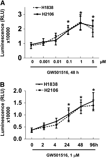
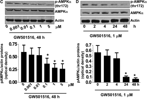
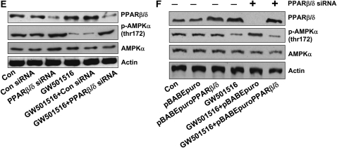
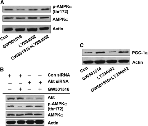
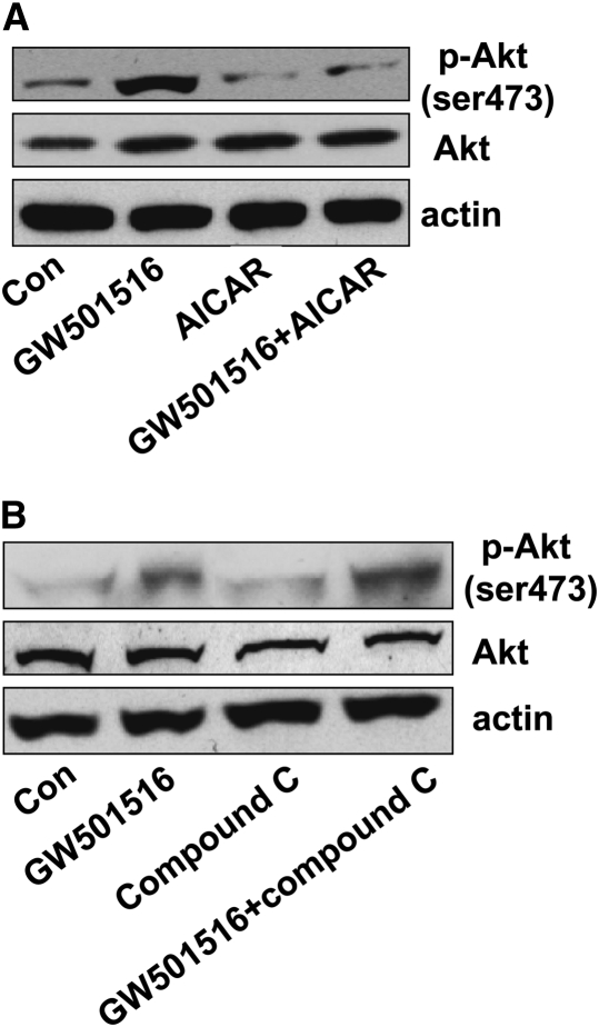
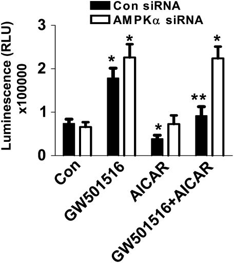
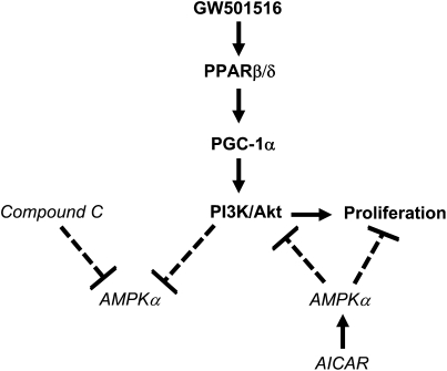
We previously demonstrated that a selective agonist of peroxisome proliferator-activated receptor beta/delta (PPARbeta/delta), GW501516, stimulated human non-small cell lung carcinoma (NSCLC) growth, partly through inhibition of phosphatase and tensin homolog deleted on chromosome 10 expression. Here, we show that GW501516 also decreases the phosphorylation of AMP-activated protein kinase alpha (AMPKalpha), a major regulator of energy metabolism. This was mediated through specific activation of PPARbeta/delta, as a PPARbeta/delta small interfering RNA inhibited the effect. However, AMPKalpha did not mediate the growth-promoting effects of GW501516, as silencing of AMPKalpha did not inhibit GW501516-induced cell proliferation. Instead, we found that GW501516 stimulated peroxisome proliferator-activated receptor coactivator gamma (PGC)-1alpha, which activated the phosphatidylinositol 3 kinase (PI3-K)/Akt mitogenic pathway. An inhibitor of PI3-K, LY294002, had no effect on PGC-1alpha, consistent with PGC-1alpha being upstream of PI3-K/Akt. Of note, an activator of AMPKalpha, 5-amino-4-imidazole carboxamide riboside, inhibited the growth-promoting effects of GW501516, suggesting that although AMPKalpha is not responsible for the mitogenic effects of GW501516, its activation can oppose these events. This study unveils a novel mechanism by which GW501516 and activation of PPARbeta/delta stimulate human lung carcinoma cell proliferation, and suggests that activation of AMPKalpha may oppose this effect.


Figures

<b>Figure 1.</b>
Figure 1.
Peroxisome proliferator–activated receptor (PPAR)-β/δ agonist increases human lung cancer cell proliferation and inhibits phosphorylation of AMP-activated protein kinase (AMPK)-α in a dose- and time-dependent manner through activation of PPARβ/δ. (A) H1838 and H2106 cells were cultured with increasing doses of GW501516 for 48 hours. Afterwards, viable cells were determined by the CellTiter-Glo Luminescent cell viability assay. Data are expressed as mean ± SD of at least three independent experiments. (B) H1838 and H2106 cells were cultured with GW501516 (1 μM) for the indicated time period. Afterwards, viable cell numbers were determined by the CellTiter-Glo luminescent cell viability assay. All data are presented as means ± SD. *Significant difference from untreated control or zero time point. (C) Cellular protein was isolated from H1838 cells cultured with increasing concentrations of GW501516 for up to 24 hours, followed by Western Blot analysis with antibodies against AMPKα and its phosphorylated form (p-AMPKα). The bar graph in the lower panel represents the mean ± SD of phospho-AMPKα/actin of at least three independent experiments. (D) Cellular protein was isolated from H1838 cells cultured with GW501516 (1 μM) for the indicated periods of time, followed by Western blot analysis with antibodies against AMPKα and its phosphorylated form (p-AMPKα). Actin served as internal control for normalization purposes. The bar graph in the lower panel represents the mean ± SD of phospho-AMPKα/actin of at least three independent experiments. *P < 0.05 when tested against control zero time point. (E) Cellular protein was isolated from H1838 cells cultured for 30 hours in the presence or absence of control or PPARβ/δ siRNA (100 nM each) before exposure of the cells to GW501516 (1 μM) for an additional 24 hours, then subjected to Western blot analysis for PPARβ/δ AMPKα and p-AMPKα. Actin served as internal control for normalization purposes. (F) Cellular protein was isolated from H1838 cells cultured for 30 hours in the presence or absence of control or PPARβ/δ siRNA (100 nM each), followed by transfection of pBABE puro and pBABEpuro PPARβ/δ plasmids (2 μg/μl each) using the Lipofectamine 2,000 reagent (Invitrogen). After 24 hours of incubation, cells were treated with or without GW501516 (1 μM) for an additional 24 hours, then subjected to Western blot analysis for PPARβ/δ AMPKα and p-AMPKα. Actin served as internal control for normalization purposes.
<b>Figure 2.</b>
Figure 2.
GW501516 increases the expression of PGC-1α protein and silencing of PGC-1α abrogates the inhibitory effect of GW501516 on phosphorylation AMPKα and Akt. (A) Cellular protein was isolated from H1838 cells cultured with increasing concentrations of GW501516 for up to 24 hours, followed by Western blot analysis with antibodies against PGC-1α. (B) Cellular protein was isolated from H1838 cells cultured with GW501516 (0.1 μM) for the indicated periods of time, followed by Western blot analysis with antibodies against PGC-1α. Actin served as internal control for normalization purposes. (C) Cellular protein was isolated from H1838 cells transfected with PGC-1α siRNA or control siRNA (100 nM each) for 40 hours before exposure of the cells to GW501516 (0.1 μM) for an additional 48 hours. Afterwards, Western blot analysis was performed using polyclonal antibodies against PGC-1α, AMPKα, and phospho-AMPKα. Actin served as internal control for normalization purposes. Con, untreated control cells. (D) Cellular protein was isolated from H1838 cells transfected with PGC-1α siRNA or control siRNA (100 nM each) for 40 hours before exposure of cells to GW501516 (0.1 μM) for an additional 2 hour. Afterwards, Western blot analysis was performed using polyclonal antibodies against PGC-1α, Akt, and phosphor-Akt (ser473). Actin served as internal control for normalization purposes.
<b>Figure 3.</b>
Figure 3.
Activation of phosphatidylinositol 3 kinase (PI3-K) is required in mediating the effect of GW501516 on phosphorylation of AMPKα. (A) Cellular protein was isolated from H1838 cells treated with LY294002 (25 μM) for 2 hours before exposure of the cells to GW501516 (1 μM) for an additional 24 hours. Afterwards, Western blot analysis was performed using polyclonal antibodies against AMPKα and phosphor-AMPKα. B, Cellular protein was isolated from H1838 cells transfected with Akt siRNA or control siRNA (100 nM each) for 40 hours before exposure of cells to GW501516 (1 μM) for an additional 48 hours. Afterwards, Western blot analysis was performed using polyclonal antibodies against Akt, AMPKα, and phosphor-AMPKα. Actin served as internal control for normalization purposes. (C) Cellular protein was isolated from H1838 cells treated with LY294002 (25 μM) for 2 hours before exposure of the cells to GW501516 (1 μM) for an additional 24 hours. Afterwards, Western blot analysis was performed using polyclonal antibodies against PGC-1α. Actin served as internal control for normalization purposes.
<b>Figure 4.</b>
Figure 4.
Activation of AMPKα blocks the stimulatory effect of GW501516 on phosphorylation of Akt. (A) Cellular protein was isolated from H1838 cells treated with 5-amino-4-imidazole carboxamide riboside (AICAR) (100 μM) for 2 hours before exposure of the cells to GW501516 (1 μM) for an additional 2 hours. Afterwards, Western blot analysis was performed using polyclonal antibodies against Akt and phosphor-Akt (ser473). Actin served as internal control for normalization purposes. (B) Cellular protein was isolated from H1838 cells treated with compound C (10 μM) for 2 hours before exposure of the cells to GW501516 (1 μM) for an additional 2 hours. Afterwards, Western blot analysis was performed using polyclonal antibodies against Akt and phosphor-Akt (ser473). Actin served as internal control for normalization purposes. Con, untreated control cells.
<b>Figure 5.</b>
Figure 5.
Activation of AMPKα inhibits GW501516-induced cell growth. H1838 cells were transfected with AMPKα siRNA or control siRNA (100 nM each) for 40 hours before exposure of cells to GW501516 (1 μM) in the presence or absence of AICAR (100 μM) for 48 hours. Afterwards, viable cell numbers were determined by the CellTiter-Glo Luminescent cell viability assay. All data are presented as means ± SD. *Significant difference from untreated control; **significance of combination treatment as compared with GW501516 alone (P < 0.05).
<b>Figure 6.</b>
Figure 6.
Schematic representation of signal pathways in response to PPARβ/δ agonist–induced NSCLC cell growth. Our findings suggest that GW501516, via activation of PPARβ/δ and the induction of PGC-1α, stimulates phosphorylation of PI3-K/Akt. This, in turn, leads to cell growth. Activation of PPARβ/δ inhibits AMPKα phosphorylation, but this event does not mediate the growth-promoting effects of GW501516. However, activation of AMPKα with AICAR inhibits PI3-K/Akt; this reduces the growth-promoting effects of GW501516. Note that the solid lines represent stimulatory effects, whereas the dotted lines represent inhibitory effects.

References

    1. Felip E, Rosell R. Testing for excision repair cross-complementing 1 in patients with non–small-cell lung cancer for chemotherapy response. Expert Rev Mol Diagn 2007;7:261–268. - PubMed
    1. Granville CA, Dennis PA. An overview of lung cancer genomics and proteomics. Am J Respir Cell Mol Biol 2005;32:169–176. - PubMed
    1. Semple RK, Chatterjee VK, O'Rahilly S. PPAR γ and human metabolic disease. J Clin Invest 2006;116:581–589. - PMC - PubMed
    1. Petrashevskaya NN, Schwarz A. Peroxisome proliferator-activated receptor β/δ: a new antihypertrophic drug target? Cardiovasc Res 2005;65:770–771. - PubMed
    1. Braissant O, Wahli W. Differential expression of peroxisome proliferator-activated receptor-α, -β, and -γ during rat embryonic development. Endocrinology 1998;139:2748–2754. - PubMed

Publication types

MeSH terms