. 2008 Oct;135(19):3247-57.
doi: 10.1242/dev.023176.

Wnt and FGF signals interact to coordinate growth with cell fate specification during limb development

Affiliations

Wnt and FGF signals interact to coordinate growth with cell fate specification during limb development

Derk ten Berge et al. Development. 2008 Oct.

Abstract

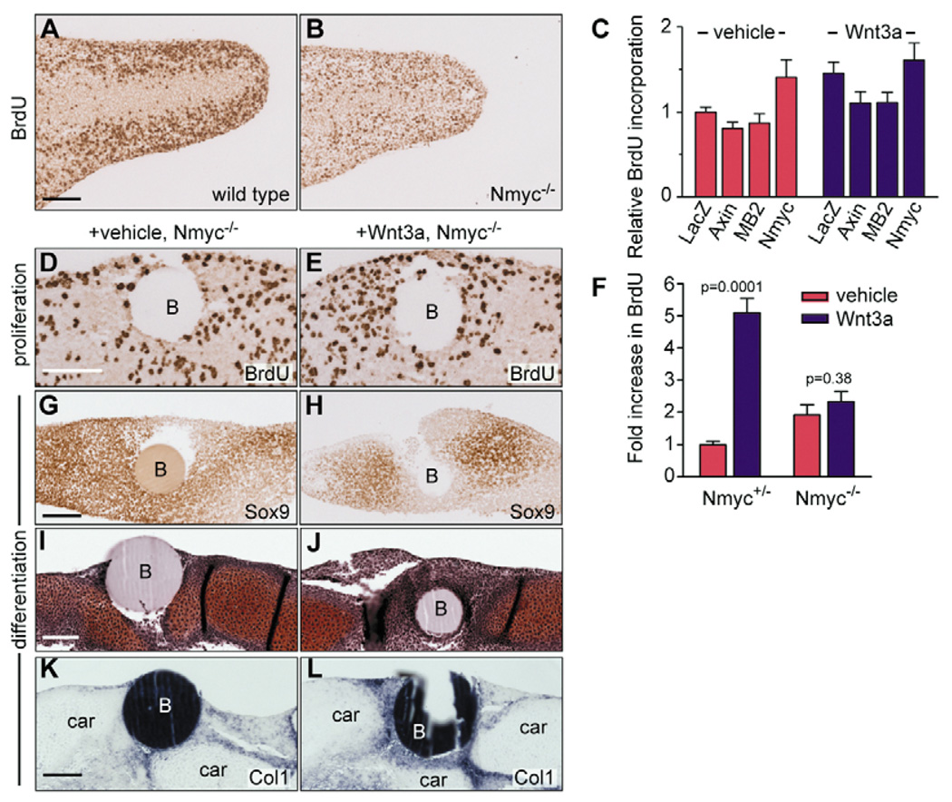
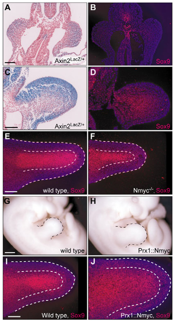
A fundamental question in developmental biology is how does an undifferentiated field of cells acquire spatial pattern and undergo coordinated differentiation? The development of the vertebrate limb is an important paradigm for understanding these processes. The skeletal and connective tissues of the developing limb all derive from a population of multipotent progenitor cells located in its distal tip. During limb outgrowth, these progenitors segregate into a chondrogenic lineage, located in the center of the limb bud, and soft connective tissue lineages located in its periphery. We report that the interplay of two families of signaling proteins, fibroblast growth factors (FGFs) and Wnts, coordinate the growth of the multipotent progenitor cells with their simultaneous segregation into these lineages. FGF and Wnt signals act together to synergistically promote proliferation while maintaining the cells in an undifferentiated, multipotent state, but act separately to determine cell lineage specification. Withdrawal of both signals results in cell cycle withdrawal and chondrogenic differentiation. Continued exposure to Wnt, however, maintains proliferation and re-specifies the cells towards the soft connective tissue lineages. We have identified target genes that are synergistically regulated by Wnts and FGFs, and show how these factors actively suppress differentiation and promote growth. Finally, we show how the spatial restriction of Wnt and FGF signals to the limb ectoderm, and to a specialized region of it, the apical ectodermal ridge, controls the distribution of cell behaviors within the growing limb, and guides the proper spatial organization of the differentiating tissues.


Figures

Fig. 1
Fig. 1. Limb ectoderm inhibits chondrogenesis and promotes proliferation via Wnt signals
(A) Chick limb ectoderm cultured on top of E11.5 Axin2lacZ/+ limb mesoderm cells induces the reporter in the mesoderm (n=10). (B) Chick limb ectoderm cultured on top of chick limb mesoderm inhibits chondrogenesis (stained with Alcian Blue) in a ~150 µm wide zone around the explant (n=30). (C) The Wnt antagonist Fz8CRD inhibits induction of the Axin2lacZ/+ reporter by the ectoderm (n=10). The ectoderm in A and C is derived from stage 24 chick limb buds and does not carry the reporter. (D) Fz8CRD protein abrogates the chondro-inhibitory effect of the ectoderm (45 out of 48). (E,F) E11.5 Axin2lacZ/+ limb bud cells induce the lacZ reporter (blue) in response to Wnt3a protein (100 ng/ml for 19 hours; F). (G,H) Chick stage 22 limb mesenchyme cells undergo chondrogenesis (stained with Alcian Blue) in micromass cultures (G), which is inhibited by 100 ng/ml Wnt3a protein (H, n>200). (I,J) Activation of the reporter by Wnt3a beads (arrowheads, J) but not vehicle beads (I) implanted in E11.5 Axin2lacZ/+ limb buds (n=4). (K,L) Vehicle beads implanted in stage 22 wing buds become embedded in the cartilage of the humerus (K), whereas chondrogenesis (stained with Alcian Blue) is inhibited around Wnt3a beads (L, n=8). (M,N) Chick limb ectoderm cultured on top of chick limb mesoderm induces BrdU incorporation (blue) in the surrounding cells (M, n=17), which is inhibited by Fz8CRD (N, n=18; nuclei labeled in red). (O,P) Sections through limb buds cultured with implanted vehicle (O) or Wnt3a (P) beads. Wnt3a induces BrdU incorporation around the bead (n=11). Scale bars: 100 µm in A–D,M–P; 500 µm in E–H; 200 µm in I–L. h, humerus; r, radius; u, ulna; B, bead; E, ectoderm.
Fig. 2
Fig. 2. Wnt3a protein re-specifies chondrogenic cells towards soft connective tissues
(A,B) Micromass cultures from chick limb mesenchyme cells treated with vehicle (A) or Wnt3a (B) for 4 days, cultured another 4 days in absence of Wnt3a. Wnt3a-treated cells failed to undergo chondrogenesis (Alcian Blue; n>50). (C) Collagen 1, tenascin C, decorin, Dermo1 and Bmp3 expression levels in high density cultures grown in continuous presence of Wnt3a (blue), or during the first 3 days of culture, after which the Wnt3a was replaced by vehicle (red). Time points 1 and 8 days were sampled twice (mean±s.e.m.). (D) Small numbers of myotubules, immunostained for myosin heavy chain (brown), accumulate in the periphery of 6-day-old mouse limb mesenchyme micromass cultures (n=6). (E) Continuous treatment with Wnt3a (250 µg/ml) slightly expands the myotubule number (n=6). (F) When Wnt3a is removed after 3 days of culture, large numbers of myotubules spread over the tissue layer (n=6). (G) Section through a chick wing 3 days after implantation of a vehicle bead at embryonic stage 22, showing the bead embedded in cartilage (n=14, Safranin O). (H) Wnt3a beads are never embedded in cartilage (n=14) but surrounded by ectopic muscle fibers, visualized by myosin heavy chain immunostaining (brown, n=7). (I,J) Section through a control chick wing (I) and a wing with implanted Wnt3a bead (J), immunostained for pro-collagen 1 (red) and myosin heavy chain (green), nuclei stained blue (DAPI) (n=4). Both sections are at a similar location and plane. Scale bars: 500 µm in A,B; 100 µm in G–J. B, bead; car, cartilage.
Fig. 3
Fig. 3. Wnt and FGF proteins act in synergy to promote proliferation and maintain the undifferentiated state
(A–G) Alcian Blue staining. (A) Fgf8 protein delays chondrogenesis in micromass cultures of chick stage 22–23 limb mesenchyme (n=12). (B) Wnt3a combined with Fgf8 blocks chondrogenesis (n=12). (C) Removal of Fgf8 at day 4 of culture has little effect on chondrogenesis (n=12). (D) Cells treated with Wnt3a and Fgf8 resume chondrogenesis upon removal of the factors at day 4 (n=12). (E–G) Limb mesenchyme cells were expanded for 4 days in presence of Wnt3a alone (E) or in combination with Fgf8 (F,G), trypsinized and replated as micromass cultures. Cells expanded in Wnt3a alone lost their chondrogenic potential (E). Cells expanded in Wnt3a and Fgf8 retained their chondrogenic potential (F), whereas Wnt3a was still able to inhibit their chondrogenesis (G) (n=4). (H) Wnt3a promotes proliferation of limb mesenchyme in micromass cultures, which is enhanced by Fgf8. Fgf8 alone does not enhance proliferation (n=4). (I) Size of limbs cultured 4 days with intact ectoderm, or without ectoderm in presence of the indicated factors (n=8, mean±s.e.m.). (J–N) Representative examples of Alcian Blue-stained limb buds cultured without ectoderm in presence of vehicle (J), Wnt3a (K), Fgf8 (L), Wnt3a and Fgf8 (M), or with ectoderm left intact (N). Scale bar: 500 µm in J–N.
Fig. 4
Fig. 4. Regulation of target genes by Wnt3a and Fgf8 in chick limb mesenchyme
Cells were cultured at high density in the presence of Wnt3a, Fgf8, or both, and samples taken 2, 4 or 6 hours after addition of the factors. Gene expression levels were plotted relative to vehicle controls. Note synergistic regulation of Nmyc, Sdc1 and Sox9 by the combination of Wnt3a and Fgf8 (blue line), and the antagonistic effect of Fgf8 on the induction of Nbl1 by Wnt3a.
Fig. 5
Fig. 5. Expression patterns of Wnt and FGF target genes correlate with cell behaviors in mouse limb buds
(A–C) Expression of Axin2lacZ/+ reporter, (D–F) BrdU labeling, (G–I) Sox9 immunostaining, (J–L) Nmyc in situ hybridization, (M) Sdc1 fluorescent immunostaining. Sections through E10.5 forelimb buds (A,D,G,J), through the level of the central metacarpal in E11.5 forelimb buds (B,E,H,K,M), through E11.5 hind limb buds (C,F,I,L). Note co-localization of proliferation (BrdU labeling, D–F) with the Axin2lacZ/+ reporter (A–C) and Nmyc expression (J–L), whereas chondrogenic differentiation (Sox9 expression, G–I) is mutually exclusive with proliferation and reporter expression. Dorsal is upwards, distal is rightwards. Scale bars: 100 µm.
Fig. 6
Fig. 6. Wnt promotes proliferation of limb mesenchyme via Nmyc and inhibits chondrogenesis independently via Sox9
(A,B) BrdU staining on longitudinal sections through the midregion of E11.5 forelimb bud of wild-type (A) and Nmyc−/− littermate (B). (C) Proliferation in E11.5 limb bud cells infected with adenovirus expressing the indicated genes (n=6). (D,E) Vehicle (D) or Wnt3a (E) beads implanted in E11.5 Nmyc−/− limb buds do not affect BrdU labeling (n=8). (F) BrdU density around vehicle or Wnt3a beads implanted in E11.5 Nmyc+/− and Nmyc−/− limbs (n=4). Wnt3a promotes proliferation 5-fold in Nmyc+/− limb buds (P=0.0001, n=4), but fails to promote proliferation in Nmyc−/− limb buds (P=0.38, n=4). (G,H) Wnt3a beads repress Sox9 in Nmyc−/− limb buds (H); vehicle beads (G) have no effect (n=4). (I,J) Wnt3a inhibits cartilage formation around the bead in Nmyc−/− limb buds (J); vehicle beads have no effect (I). Cartilage stained red with Safranin O (n=4). (K,L) Wnt3a beads (L), but not vehicle beads (K), induce collagen 1 in Nmyc−/− limb buds (n=4). Dorsal is upwards, distal rightwards (A,B). Scale bars: 100 µm. B, bead; car, cartilage.
Fig. 7
Fig. 7. Wnt couples expansion to differentiation
(A) Axin2lacZ expression (blue) is visible throughout the forelimb buds of E9.5 embryos (22 somites). (B) Sox9 expression (red) is absent in a nearby section through the same embryo as in A. Expression of Sox9 can be seen in other areas of the embryo, such as the neural tube. (C,D) At late E9.5 (27 somites), Axin2lacZ is no longer active in the centre of the forelimb (C), where Sox9 is now expressed (D). (E,F) Sox9 immunostaining on longitudinal sections through the midregion of stage-matched wild-type (E) and Nmyc−/− (F) E11.5 forelimbs. (G,H) Stage-matched E11.5 wild type (G) and Prx1::Nmyc embryo (H, n=5). (I,J) Sox9 immunostaining on longitudinal sections through the midregion of the forelimbs of the embryos shown in G,H. Dorsal is upwards and distal is rightwards (C–F,I,J). Nuclei are labeled in blue (B,D,E,F,I,J). Scale bars: 100 µm in A-F,I,J; 500 µm in G,H.
Fig. 8
Fig. 8. Wnt and FGF signals interact to coordinate growth and cell fate specification during limb development
In the newly established limb bud (E9.5), both Wnt and FGF proteins signal throughout the limb mesenchyme and maintain all cells in a multipotent, proliferative state (indicated by red/blue hatching, marked by Axin2 and Dusp6). Following limb outgrowth, cells in the center of the limb are no longer within range of the signals. This allows cell cycle withdrawal and expression of Sox9, leading to establishment of the chondrogenic core (indicated in blue, marked by Sox9). In the periphery, meanwhile, cells out of range of FGFs from the AER are still within range of Wnts from the ectoderm (indicated by red hatching, marked by Nbl1), which maintains the proliferative state at a lower level, and respecifies the cells towards soft connective tissue fates. As a result of these processes, a proximodistally extended organ forms with a multipotent, rapidly growing tip and a chondrogenic core surrounded by soft connective tissues.

Similar articles

Cited by

References

    1. Abe G, Ide H, Tamura K. Function of FGF signaling in the developmental process of the median fin fold in zebrafish. Dev. Biol. 2007;304:355–366. - PubMed
    1. Ahrens PB, Solursh M, Reiter RS. Stage-related capacity for limb chondrogenesis in cell culture. Dev. Biol. 1977;60:69–82. - PubMed
    1. Akiyama H, Chaboissier M-C, Martin JF, Schedl A, de Crombrugghe B. The transcription factor Sox9 has essential roles in successive steps of the chondrocyte differentiation pathway and is required for expression of Sox5 and Sox6. Genes Dev. 2002;16:2813–2828. - PMC - PubMed
    1. Anakwe K, Robson L, Hadley J, Buxton P, Church V, Allen S, Hartmann C, Harfe B, Nohno T, Brown AM, et al. Wnt signalling regulates myogenic differentiation in the developing avian wing. Development. 2003;130:3503–3514. - PubMed
    1. Aulehla A, Wehrle C, Brand-Saberi B, Kemler R, Gossler A, Kanzler B, Herrmann BG. Wnt3a plays a major role in the segmentation clock controlling somitogenesis. Dev. Cell. 2003;4:395–406. - PubMed

Publication types