. 2008;110(2):e49-58.
doi: 10.1159/000153245. Epub 2008 Sep 8.

Expression of ammonia transporters, Rhbg and Rhcg, in chronic cyclosporine nephropathy in rats

Affiliations

Expression of ammonia transporters, Rhbg and Rhcg, in chronic cyclosporine nephropathy in rats

Sun Woo Lim et al. Nephron Exp Nephrol. 2008.

Abstract

Background/aims: Cyclosporine (CsA)-induced renal injury causes renal tubular acidosis. The current study was performed to evaluate the influence of CsA-induced renal injury on the ammonia transporter family members, Rh B-glycoprotein (Rhbg) and Rh C-glycoprotein (Rhcg).

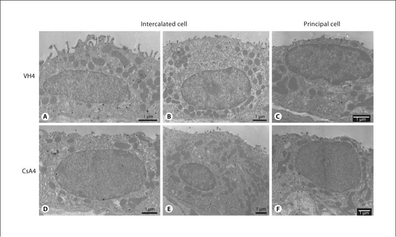
Methods: Rats were treated daily for 1 or 4 weeks with vehicle (VH) or CsA. Induction of chronic CsA-induced nephropathy was confirmed by demonstrating impaired renal function and characteristic histopathology. Rhbg and Rhcg expression was evaluated with immunoblot, immunohistochemistry, real-time RT-PCR and electron microscopy.

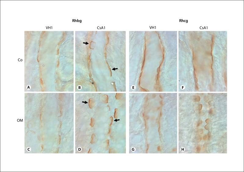
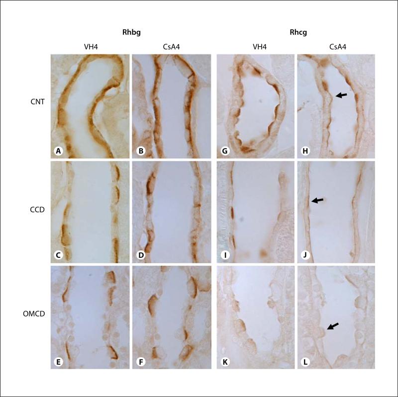
Results: CsA treatment for 4 weeks developed mild metabolic acidosis and decreased urinary ammonia excretion. Rhcg mRNA expression was unchanged in both the cortex and outer medulla, but Rhcg protein expression in the CsA group was significantly reduced in the cortex and outer medulla. There were no significant differences in Rhbg mRNA and protein expression between the CsA and VH group.

Conclusion: Long-term treatment with CsA in rats results in decreased urinary ammonia excretion accompanied by decreased expression of Rhcg; these changes are likely to mediate the CsA-induced defect in ammonium excretion in the collecting duct.


Figures

Fig. 1
Fig. 1
Western blot and densitometric analysis of Rhbg (A, C) and Rhcg (B, D) in the cortex (Co) and outer medulla (OM) of VH- (□)) or CsA-treated rat kidneys (■). Each lane was loaded with a sample from a different animal. Both of Rhbg and Rhcg are prominent with a size of 50 kDa. At 1 week, there was a significant increase in the protein level of Rhbg in the CsA group compared with the VH group. But Rhcg was not changed significantly between the 2 groups. At 4 weeks, there was no significant difference in the protein level of Rhbg between the experimental groups. However, the amount of Rhcg significantly decreased in the CsA group treated for 4 weeks compared with the VH group treated for 4 weeks. VH1 = Vehicle group treated for 1 week; CsA1 = cyclosporine group treated for 1 week; VH4 = vehicle group treated for 4 weeks; CsA4 = cyclosporine group treated for 4 weeks. Values are referred to VH as 100%. * p < 0.05 vs. VH.
Fig. 2
Fig. 2
Representative micrographs of immunohistochemistry for Rhbg (A–D) and Rhcg (E–H) in the cortical collecting duct (CCD; A, B, E, F), outer medullary collecting duct (OMCD; C, D, G, H) of rat kidneys treat with VH (A, C, E, G) or CsA (B, D, F, and H) for 1 week. Predominant immunoreactivity for Rhbg was detected in the basolateral plasma membrane of the cortical collecting duct (CCD) and outer medullary collecting duct (OMCD). Rhbg expression was increased in rat kidneys treated with CsA for 1 week compared with VH-treated rat kidneys (A–D). Intense immuno-reactivity for Rhcg was detected in the apical plasma membrane of CCD and OMCD. Rhcg expression did not alter in CsA rat kidneys treated with CsA for 1 week compared with VH-treated rat kidneys (E–H). Arrows indicate a strong positive Rhbg reaction from the apical side of the collecting duct. Co = Cortex; OM = outer medulla. Magnification × 1,000.
Fig. 3
Fig. 3
Representative micrographs of immunohistochemistry for Rhbg (A–F) and Rhcg (G–L) in the connecting tubule (CNT; A, B, G, H), cortical collecting duct (CCD; C, D, I, J), and outer medullary collecting duct (OMCD; E, F, K, L) of rat kidneys treated with VH (A, C, E, G, I, K) or CsA (B, D, F, H, J, L) for 4 weeks. Predominant immunoreactivity for Rhbg was detected in the basolateral plasma membrane of CNT, CCD, and OMCD. Rhbg expression did not alter in rat kidneys treat with CsA for 4 weeks compared with VH-treated rat kidneys (A–F). Intense immunoreactivity for Rhcg was detected in the apical plasma membrane of CNT, CCD, and OMCD. Rhcg expression was reduced in those regions of the rat kidney treated with CsA for 4 weeks compared with VH-treated rat kidney (G–L). Arrows indicate a weak positive Rhcg reaction from the apical side of the collecting duct. VH4 = Vehicle group treated for 4 weeks; CsA4 = cyclosporine group treated for 4 weeks. Magnification × 1,000.
Fig. 4
Fig. 4
Transmission electron microscopic localization of Rhcg in the intercalated cells (A, B, D, E) and principal cells (C, F) from the cortical collecting duct of rat kidneys treated with VH (A–C) and CsA (D–F) for 4 weeks using an immunogold method. In the VH-treated group, numerous amounts of Rhcg immunolabeling are shown in the apical membrane microprojection, subapical region, and basal membrane (A), whereas Rhcg labeling is lower the CsA-treated group with short apical microprojections (D). Rhcg immunolabeling is mainly present in the apical region in type B intercalated cell and principal cell. However, there is no difference in Rhcg labeling in between VH- and CsA-treated groups (B, C, E, F).
Fig. 5
Fig. 5
mRNA expression of Rhbg and Rhcg using real-time PCR in the cortex and outer medulla (OM) from VH- (□)) or CsA-treated rat kidneys (■) for 1 week (A) and 4 weeks (B). CsA treatment did not alter mRNA expression of both Rhbg and Rhcg from cortex and OM. Relative Rhbg or Rhcg mRNA values are normalized to GAPDH mRNA. NS = No significance.

Similar articles

Cited by

References

    1. Ling BN, Eaton DC. Cyclosporin A inhibits apical secretory K + channels in rabbit cortical collecting tubule principal cells. Kidney Int. 1993;44:974–984. - PubMed
    1. Thompson CB, June CH, Sullivan KM, Thomas ED. Association between cyclosporin neurotoxicity and hypomagnesaemia. Lancet. 1984;17:1116–1120. - PubMed
    1. Young BA, Burdmann EA, Johnson RJ, Alpers CE, Giachelli CM, Eng E, Andoh T, Bennett WM, Couser WG. Cellular proliferation and macrophage influx precede interstitial fibrosis in cyclosporine nephrotoxicity. Kidney Int. 1995;48:439–448. - PubMed
    1. Batlle DC, Gutterman C, Tarka J, Prasad R. Effect of short-term cyclosporine A administration on urinary acidification. Clin Nephrol. 1986;25(suppl 1):S62–S69. - PubMed
    1. DuBose TD, Jr, Good DW, Hamm LL, Wall SM. Ammonium transport in the kidney: new physiological concepts and their clinical implications. J Am Soc Nephrol. 1991;1:1193–1203. - PubMed

Publication types

MeSH terms