. 2008 Oct;9(10):1131-9.
doi: 10.1038/ni.1652. Epub 2008 Sep 7.

ThPOK acts late in specification of the helper T cell lineage and suppresses Runx-mediated commitment to the cytotoxic T cell lineage

Affiliations

ThPOK acts late in specification of the helper T cell lineage and suppresses Runx-mediated commitment to the cytotoxic T cell lineage

Takeshi Egawa et al. Nat Immunol. 2008 Oct.

Abstract

The transcription factor ThPOK is required and sufficient for the generation of CD4(+)CD8(-) thymocytes, yet the mechanism by which ThPOK orchestrates differentiation into the CD4(+) helper T cell lineage remains unclear. Here we used reporter mice to track the expression of transcription factors in developing thymocytes. Distal promoter-driven expression of the gene encoding the transcription factor Runx3 was restricted to major histocompatibility complex (MHC) class I-selected thymocytes. In ThPOK-deficient mice, such expression was derepressed in MHC class II-selected thymocytes, which contributed to their redirection to the CD8(+) T cell lineage. In the absence of both ThPOK and Runx, redirection was prevented and cells potentially belonging to the CD4(+) lineage, presumably specified independently of ThPOK, were generated. Our results suggest that MHC class II-selected thymocytes are directed toward the CD4(+) lineage independently of ThPOK but require ThPOK to prevent Runx-dependent differentiation toward the CD8(+) lineage.


Figures

Figure 1
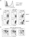
Figure 1. CD8SP lineage-specific Runx3 expression from its distal promoter is required for Cd4 silencing and CD103 expression in CD8+ T cells
(a) Runx3 and Runx1 protein expression in purified CD8+ cells from Runx3dYFP/YFP and Runx3dYFP/+ mice is shown with anti-HMG1 blot as loading control. Lysates from Cd4-cre+Runx1F/F CD4+ T cells and Cd4-cre+Runx3F/F CD8+ T cells were used as negative controls for Runx1 and Runx3 expression, respectively. (b,c) CD4 and CD8 (b) or CD8 and CD103 (c) expression in TCRβ+ lymph node (LN) cells from Runx3dYFP/YFP and Runx3dYFP/+ mice. In the right panel in (b), CD4 expression in CD8+ T cells from Runx3dYFP/YFP (open histogram) and Runx3dYFP/+ (shaded histogram) mice is shown. Mean fluorescent intensity: Runx3dYFP/+ 308, Runx3dYFP/YFP 1767. Data shown are representative of more than 3 independent experiments.
Figure 2
Figure 2. Runx3d and ThPOK reporter expression in developing thymocytes
(a) Distal promoter-derived Runx3 expression determined by the Runx3d-YFP reporter in thymocyte subpopulations as defined in Supplementary Fig. 2, online. YFP+ cells are gated with orange rectangles. (b) ThPOK-GFP reporter expression in developing thymocytes. GFPhi and GFPlo populations are gated with dark and light blue rectangles, respectively. Percentages indicate frequencies of YFP+, GFPhi, and GFPlo cells among thymocyte subsets marked at top, except for those in the CD4+CD8+ subpopulation, for which percentages correspond to frequencies of gated cells among CD4+CD8+TCRβhi thymocytes (marked with asterisks). (c) ThPOK-GFP reporter expression in MHCI- and MHCII-restricted CD4+CD8lo thymocyte subpopulations. The percentages of total GFP+ cells (bottom gate) and GFPhi cells (top gate) are shown. (d) Mutually exclusive high ThPOK-GFP and Runx3d-YFP expression during thymocyte differentiation. ThPOK-GFP and Runx3d-YFP expression in developing thymocytes from indicated mice. Numbers indicate percentages of cells within gates. (e) Continued ThPOK-GFP expression in redirected MHCII-restricted ThPOK-deficient CD8+ T cells. ThPOK-GFP expression in CD8+ T cells from Zbtb7bGFP/+ or Zbtb7bGFP/− mice. Numbers indicate percentages of GFP+ cells. Data shown are representative of more than 3 independent experiments.
Figure 3
Figure 3. CD8+ lineage-specific Runx3d-YFP expression is de-repressed in ThPOK-deficient MHCII-restricted thymocytes
(a) ThPOK up-regulation independently of ThPOK following positive selection. ThPOK-GFP reporter expression in HSAhiCD69+CD4+CD8lo/− thymocytes from Zbtb7b+/+, Zbtb7bGFP/+ and Zbtb7bGFP/− mice. (b) Runx3d-YFP expression in thymocyte subpopulations in the presence or absence of ThPOK. YFP+ cells are gated as indicated by rectangles. (c) CD8 down-regulation following thymocyte positive selection in the absence of ThPOK. CD4 and CD8 expression in HSAhiCD69+ thymocytes from Zbtb7bGFP/+, Zbtb7bGFP/− and H2-Ab1−/− mice is shown. CD4+CD8 cells are gated as shown within rectangles and their frequencies are indicated. Data shown are representative of more than 3 independent experiments.
Figure 4
Figure 4. CD4+ T cell differentiation in the presence of reduced amounts of ThPOK
(a,b) ThPOK expression in CD4+CD8 T cells from Zbtb7bFN/− mice. ThPOK expression was examined by qRT-PCR (a) and immunoblotting (b). qRT-PCR data are shown as averages and standard deviations from three independent samples. (c) Lineage redirection of MHCII-restricted T cells to the CD8 lineage in the presence of reduced amounts of ThPOK. CD4 and CD8 expression in gated HSAlo/−TCRβhi mature thymocytes from Zbtb7b+/−, Zbtb7bFN/−, Zbtb7b+/−B2m−/− and Zbtb7bFN/−B2m−/− mice is shown. Percentages of CD4SP, CD8SP, CD4+CD8+ and CD4CD8 cells are indicated. (d) De-repressed Runx3 protein expression in CD4+CD8 T cells from Zbtb7bFN/− mice. Runx1 and Runx3 expression (indicated by arrows) was examined by immunoblotting with anti-pan-Runx. Anti-HMG1 was used as loading control. Data shown are representative of more than 3 independent experiments.
Figure 5
Figure 5. ThPOK is dispensable for differentiation of CD4SP thymocytes
(a) HSA and TCRβ expression in total thymocytes (top), and CD4 and CD8 expression in HSAlo/− TCRβhi mature thymocytes (middle) and TCRβ+B220 peripheral T cells (bottom) in the absence of ThPOK, CBFβ, or both. The CD4CD8+ cells shown with asterisks in Lck-cre+CbfbF/F and Lck-cre+ CbfbF/FZbtb7bGFP/− panels are those that escaped Cre-mediated inactivation of Cbfb and hence have normal Cd4 silencing. Data shown here are representative from two independent experiments with similar results. (b) Absolute numbers of HSA+CD69+ positively selected thymocytes and HSAlo/− mature CD4SP thymocytes in the absence of ThPOK, CBFβ, or both. Each column shows cell numbers in a mouse with each of the genotypes.
Figure 6
Figure 6. De-repression of CD8+ lineage-specific genes in MHCII-restricted cells in the absence of ThPOK or in the presence of insufficient amount of ThPOK
(a) MHCII-restricted HSAhiCD69+CD4+CD8lo/− ThPOK-GFPhi thymocytes from Zbtb7bGFP/+ (black columns) or Zbtb7bGFP/− (white columns) were purified using gates as shown in Fig. 3, and expression of CD4+ lineage-specific genes (Cd4, St8si6, Zbtb7b and Gata3) and CD8+ lineage-specific genes (Cd8a, Itgae, Prf1, Gpr114, Nkg7 and Cd160) was examined by qRT-PCR. mRNA expression of individual genes was normalized against Hprt1 expression and average expression in Zbtb7bGFP/+ cells was set as 1. (b) Intracellular staining for IFN-γ and IL-4 in Zbtb7bFN/− CD4+CD8 T cells following 3 days of stimulation with anti-CD3 and anti-CD28 in the absence of IL-12. (c) Expression of the CD8+ lineage-specific IFN-γ regulator Eomes in CD4+ T cells from wild-type and Zbtb7bFN/− mice and in wild-type CD8+ T cells, as quantified by q-RT-PCR. Hprt1-normalized Eomes mRNA expression is shown as average and standard deviations from three independent samples. Statistical difference was tested by two-tailed T test with assumption of unequal variance. Genes that showed a P value smaller than 0.05 are marked with asterisks in (a). Actual P values for individual genes were as follows: Cd4: 0.28, St8sia6: 0.13, Zbtb7b: 0.01, Gata3: 0.5, Cd8a: 0.04, Itgae: 0.04, Prf1: 0.01, Gpr114: 0.09, Nkg7: 0.04, Cd160: 0.02.

Comment in

Similar articles

Cited by

References

    1. Kappes DJ, He X, He X. CD4-CD8 lineage commitment: an inside view. Nat Immunol. 2005;6:761–766. - PubMed
    1. Zou YR, et al. Epigenetic silencing of CD4 in T cells committed to the cytotoxic lineage. Nat Genet. 2001;29:332–336. - PubMed
    1. Sawada S, Scarborough JD, Killeen N, Littman DR. A lineage-specific transcriptional silencer regulates CD4 gene expression during T lymphocyte development. Cell. 1994;77:917–929. - PubMed
    1. Kioussis D, Ellmeier W. Chromatin and CD4, CD8A and CD8B gene expression during thymic differentiation. Nat Rev Immunol. 2002;2:909–919. - PubMed
    1. Laslo P, et al. Multilineage transcriptional priming and determination of alternate hematopoietic cell fates. Cell. 2006;126:755–766. - PubMed

Publication types

MeSH terms

Substances