

Identification of candidate genes for human retinal degeneration loci using differentially expressed genes from mouse photoreceptor dystrophy models
- PMID: 18776951
- PMCID: PMC2529471
Identification of candidate genes for human retinal degeneration loci using differentially expressed genes from mouse photoreceptor dystrophy models
Abstract
Purpose: Retinal degeneration (RD) is a complex mechanism that appears to involve many biologic processes including oxidative stress, apoptosis, and cellular remodeling. Currently there are 51 mapped, but not identified, RD human disease loci.
Methods: To assign possible disease genes to RD loci, we have used a comparative genomics procedure that incorporates microarray gene expression data of three independent mouse models for photoreceptor dystrophy (rd1, rd2, and constant light-damage in BALB/c mice), human ortholog data, and databases of known chromosomal locations involved in human RD. Immunohistochemistry and enzyme activity assays were used to further characterize a candidate gene product.
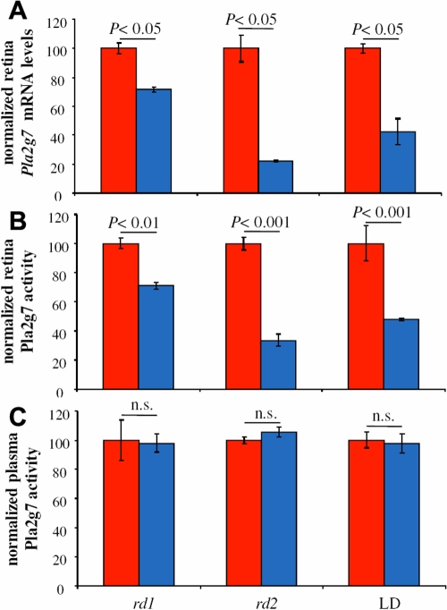
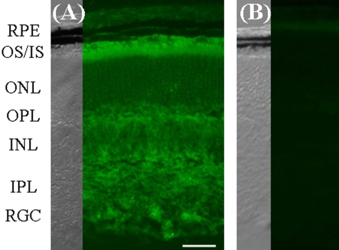
Results: Our analysis yielded candidate genes for four mapped, but unsolved, human chromosomal locations and confirmed two previously identified monogenic disease loci for human RD, thus validating our approach. PLA2G7 (phospholipase A2, group VII; PAF-AH, Lp-PLA2), a candidate for a dominant form macular dystrophy (Benign Concentric Annular Macular Dystrophy [BCMAD]), was selected for further study. The PLA2G7 enzyme is known to mediate breakdown of oxidatively damaged phospholipids, a contributor to oxidative stress in the retina. PLA2G7 protein was enriched in mouse photoreceptor inner and outer segments. In the rd1, rd2, and BALB/c mice exposed to constant light, retinal tissue activity levels, but not plasma levels, were significantly reduced at the onset of photoreceptor cell death.
Conclusions: We have shown that this comparative genomics approach verified existing RD genes as well as identified novel RD candidate genes. The results on the characterization of the PLA2G7 protein, one of the novel RD genes, suggests that retinal tissue PLA2G7 levels may constitute an important risk factor for BCMAD. In summary, this reverse mapping approach, using accepted mouse models of human disease and known human RD loci, may prove useful in identifying possible novel disease candidates for RD and may be applicable to other human diseases.
Figures
Similar articles
-
Multiple, parallel cellular suicide mechanisms participate in photoreceptor cell death.Exp Eye Res. 2006 Aug;83(2):380-9. doi: 10.1016/j.exer.2006.01.014. Epub 2006 Apr 19. Exp Eye Res. 2006. PMID: 16626700
-
Retinal degeneration in the rd mouse in the absence of c-fos.Invest Ophthalmol Vis Sci. 1998 Nov;39(12):2239-44. Invest Ophthalmol Vis Sci. 1998. PMID: 9804131
-
Strain difference in photoreceptor cell death after retinal detachment in mice.Invest Ophthalmol Vis Sci. 2014 May 22;55(7):4165-74. doi: 10.1167/iovs.14-14238. Invest Ophthalmol Vis Sci. 2014. PMID: 24854853 Free PMC article.
-
Retinal degeneration mutants in the mouse.Vision Res. 2002 Feb;42(4):517-25. doi: 10.1016/s0042-6989(01)00146-8. Vision Res. 2002. PMID: 11853768 Review.
-
Photoreceptor cell death mechanisms in inherited retinal degeneration.Mol Neurobiol. 2008 Dec;38(3):253-69. doi: 10.1007/s12035-008-8045-9. Epub 2008 Nov 4. Mol Neurobiol. 2008. PMID: 18982459 Review.
Cited by
-
Macrophage VLDL receptor promotes PAFAH secretion in mother's milk and suppresses systemic inflammation in nursing neonates.Nat Commun. 2012;3:1008. doi: 10.1038/ncomms2011. Nat Commun. 2012. PMID: 22910354 Free PMC article.
-
Intravitreal dobesilate in the treatment of choroidal neovascularisation associated with age-related macular degeneration: report of two cases.BMJ Case Rep. 2012 Sep 3;2012:bcr2012006619. doi: 10.1136/bcr-2012-006619. BMJ Case Rep. 2012. PMID: 22948997 Free PMC article.
-
Loss of Otx2 in the adult retina disrupts retinal pigment epithelium function, causing photoreceptor degeneration.J Neurosci. 2013 Jun 12;33(24):9890-904. doi: 10.1523/JNEUROSCI.1099-13.2013. J Neurosci. 2013. PMID: 23761884 Free PMC article.
-
A global transcriptome analysis reveals molecular hallmarks of neural stem cell death, survival, and differentiation in response to partial FGF-2 and EGF deprivation.PLoS One. 2013;8(1):e53594. doi: 10.1371/journal.pone.0053594. Epub 2013 Jan 7. PLoS One. 2013. PMID: 23308259 Free PMC article.
-
Changes in gene expression associated with retinal degeneration in the rd3 mouse.Mol Vis. 2013 May 6;19:955-69. Print 2013. Mol Vis. 2013. PMID: 23687432 Free PMC article.
References
-
- Koenekoop RK, Lopez I, den Hollander AI, Allikmets R, Cremers FP. Genetic testing for retinal dystrophies and dysfunctions: benefits, dilemmas and solutions. Clin Experiment Ophthalmol. 2007;35:473–85. - PubMed
-
- Farber DB. From mice to men: the cyclic GMP phosphodiesterase gene in vision and disease. The Proctor Lecture. Invest Ophthalmol Vis Sci. 1995;36:263–75. - PubMed
-
- Travis GH, Brennan MB, Danielson PE, Kozak CA, Sutcliffe JG. Identification of a photoreceptor-specific mRNA encoded by the gene responsible for retinal degeneration slow (rds). Nature. 1989;338:70–3. - PubMed
Publication types
MeSH terms
Substances
Grants and funding
LinkOut - more resources
Full Text Sources
Other Literature Sources
Molecular Biology Databases
Miscellaneous