

An automated CT based lung nodule detection scheme using geometric analysis of signed distance field
- PMID: 18777905
- PMCID: PMC2673651
- DOI: 10.1118/1.2948349
An automated CT based lung nodule detection scheme using geometric analysis of signed distance field
Abstract
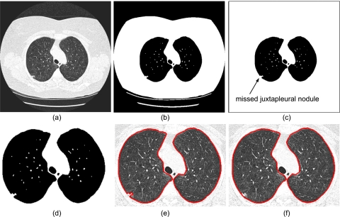
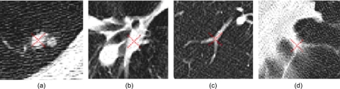
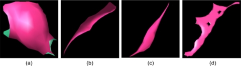
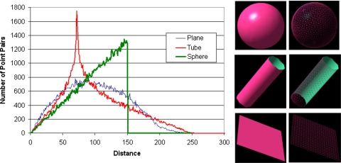
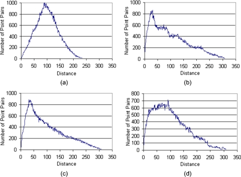
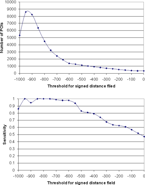
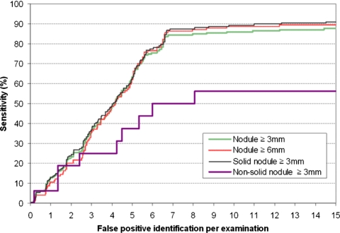
The authors present a new computerized scheme to automatically detect lung nodules depicted on computed tomography (CT) images. The procedure is performed in the signed distance field of the CT images. To obtain an accurate signed distance field, CT images are first interpolated linearly along the axial direction to form an isotropic data set. Then a lung segmentation strategy is applied to smooth the lung border aiming to include as many juxtapleural nodules as possible while minimizing over segmentations of the lung regions. Potential nodule regions are then detected by locating local maximas of signed distances in each subvolume with values and the sizes larger than the smallest nodule of interest in the three-dimensional space. Finally, all detected candidates are scored by computing the similarity distance of their medial axis-like shapes obtained through a progressive clustering strategy combined with a marching cube algorithm from a sphere based shape. A free-response receiver operating characteristics curve is computed to assess the scheme performance. A performance test on 52 low-dose CT screening examinations that depict 184 verified lung nodules showed that during the initial stage the scheme achieved an asymptotic maximum sensitivity of 95.1% (175/184) with an average of 1200 suspicious voxels per CT examination. The nine missed nodules included two small solid nodules (with a diameter < or =3.1 mm) and seven nonsolid nodules. The final performance level after the similarity scoring stage was an absolute sensitivity level, namely, including the nine missed during the initial stage, of 81.5% (150/184) with 6.5 false-positive identifications per CT examination. This preliminary study demonstrates the feasibility of applying a simple and robust geometric model using the signed distance field to identify suspicious lung nodules. In the authors' data set the sensitivity of this scheme is not affected by nodule size. In addition to potentially being a stand alone approach, the signed distance field based method can be easily implemented as an initial filtering step in other computer-aided detection schemes.
Figures

Similar articles
-
Refinement of lung nodule candidates based on local geometric shape analysis and Laplacian of Gaussian kernels.Comput Biol Med. 2014 Nov;54:188-98. doi: 10.1016/j.compbiomed.2014.09.010. Epub 2014 Sep 28. Comput Biol Med. 2014. PMID: 25303113
-
High performance lung nodule detection schemes in CT using local and global information.Med Phys. 2012 Aug;39(8):5157-68. doi: 10.1118/1.4737109. Med Phys. 2012. PMID: 22894441 Free PMC article.
-
Computer-aided diagnostic scheme for the detection of lung nodules on chest radiographs: localized search method based on anatomical classification.Med Phys. 2006 Jul;33(7):2642-53. doi: 10.1118/1.2208739. Med Phys. 2006. PMID: 16898468
-
Computerized detection of lung nodules in thin-section CT images by use of selective enhancement filters and an automated rule-based classifier.Acad Radiol. 2008 Feb;15(2):165-75. doi: 10.1016/j.acra.2007.09.018. Acad Radiol. 2008. PMID: 18206615 Free PMC article.
-
A novel lung nodules detection scheme based on vessel segmentation on CT images.Biomed Mater Eng. 2014;24(6):3179-86. doi: 10.3233/BME-141139. Biomed Mater Eng. 2014. PMID: 25227026
Cited by
-
Multi-Modal Feature Fusion-Based Multi-Branch Classification Network for Pulmonary Nodule Malignancy Suspiciousness Diagnosis.J Digit Imaging. 2023 Apr;36(2):617-626. doi: 10.1007/s10278-022-00747-z. Epub 2022 Dec 7. J Digit Imaging. 2023. PMID: 36478311 Free PMC article.
-
Machine Learning in Computer-aided Diagnosis of the Thorax and Colon in CT: A Survey.IEICE Trans Inf Syst. 2013 Apr 1;E96-D(4):772-783. doi: 10.1587/transinf.e96.d.772. IEICE Trans Inf Syst. 2013. PMID: 24174708 Free PMC article.
-
CT Quantitative Analysis and Its Relationship with Clinical Features for Assessing the Severity of Patients with COVID-19.Korean J Radiol. 2020 Jul;21(7):859-868. doi: 10.3348/kjr.2020.0293. Korean J Radiol. 2020. PMID: 32524786 Free PMC article.
-
Vasculature surrounding a nodule: A novel lung cancer biomarker.Lung Cancer. 2017 Dec;114:38-43. doi: 10.1016/j.lungcan.2017.10.008. Epub 2017 Oct 27. Lung Cancer. 2017. PMID: 29173763 Free PMC article.
-
Computer-aided diagnosis systems for lung cancer: challenges and methodologies.Int J Biomed Imaging. 2013;2013:942353. doi: 10.1155/2013/942353. Epub 2013 Jan 29. Int J Biomed Imaging. 2013. PMID: 23431282 Free PMC article.
References
-
- Kaneko M., Eguchi K., Ohmatsu H., Naruke T., Suemasu K., and Moriyama N., “Peripheral lung cancer: Screening and detection with low-dose spiral CT versus radiography,” Radiology 201, 798–802 (1996). - PubMed
-
- Diederich S., Lenzen H., Windmann R., Puskas R. Z., Yelbuz T. M., Henneken S., Klaiber T., Eameri M., Roos N., and Peters P. E., “Pulmonary nodules: Experimental and clinical studies at low-dose CT,” Radiology 213, 289–298 (1999). - PubMed
-
- Detailed guide: What are the key statistics about lung cancer, http://www.cancer.org/docroot/, revised on October 15, 2007.
-
- Fischbach F., Knollmann F., Griesshaber V., Freund T., Akkol E., and Felix R., “Detection of pulmonary nodules by multislice computed tomography: Improved detection rate with reduced slice thickness,” Eur. Radiol. 13, 2378–2383 (2003). - PubMed
Publication types
MeSH terms
Grants and funding
LinkOut - more resources
Full Text Sources
Medical
