. 2008 Aug;35(8):3462-70.
doi: 10.1118/1.2948388.

Effects of variation in perfusion rates and of perfusion models in computational models of radio frequency tumor ablation

Affiliations

Effects of variation in perfusion rates and of perfusion models in computational models of radio frequency tumor ablation

David J Schutt et al. Med Phys. 2008 Aug.

Abstract

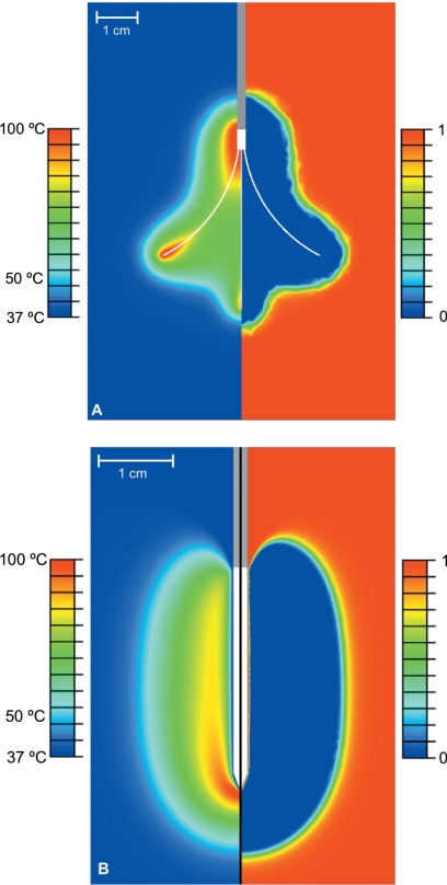
Purpose: Finite element method (FEM) models are commonly used to simulate radio frequency (RF) tumor ablation. Prior FEM models of RF ablation have either ignored the temperature dependent effect of microvascular perfusion, or implemented the effect using simplified algorithms to reduce computational complexity. In this FEM modeling study, the authors compared the effect of different microvascular perfusion algorithms on ablation zone dimensions with two commercial RF electrodes in hepatic tissue. They also examine the effect of tissue type and inter-patient variation of perfusion on ablation zone dimensions.

Methods and materials: The authors created FEM models of an internally cooled and multi-tined expandable electrode. RF voltage was applied to both electrodes (for 12 or 15 min, respectively) such that the maximum temperature in the model was 105 degrees C. Temperature dependent microvascular perfusion was implemented using three previously reported methodologies: cessation above 60 degrees C, a standard first-order Arrhenius model with decreasing perfusion with increasing degree of vascular stasis, and an Arrhenius model that included the effects of increasing perfusion at the ablation zone boundary due to hyperemia. To examine the effects of interpatient variation, simulations were performed with base line and +/-1 standard deviation values of perfusion. The base line perfusion was also varied to simulate the difference between normal and cirrhotic liver tissue.

Results: The ablation zone volumes with the cessation above 60 degrees C perfusion algorithm and with the more complex Arrhenius model were up to 70% and 25% smaller, respectively, compared to the standard Arrhenius model. Ablation zone volumes were up to 175% and approximately 100% different between the simulations where -1 and +1 standard deviation values of perfusion were used in normal and cirrhotic liver tissue, respectively.

Conclusions: The choice of microvascular perfusion algorithm has significant effects on final ablation zone dimensions in FEM models of RF ablation. The authors also found that both interpatient variation in base line tissue perfusion and the reduction in perfusion due to cirrhosis have considerable effect on ablation zone dimensions.


Figures

Figure 1
Figure 1
Fully deployed Rita Starburst XL electrode used in FEM. The prongs and the distal 5 mm of the shaft are electrically conductive.
Figure 2
Figure 2
Close-up of mesh around the tip of the internally cooled (a) and multi-tined expandable electrodes (b). The node spacing at the boundary between the tissue (white) and the electrodes (gray) was 0.1 mm.
Figure 3
Figure 3
Relative perfusion vs degree of stasis (DS) used in algorithm 2 and 3. For algorithm 3 (solid line), the graph depicts the original curve determined experimentally in renal tissue in He et al. (Ref. 30), which was modeled as a series of linear approximations. For algorithm 2 (dotted line), the relative perfusion was inversely proportional to DS.
Figure 4
Figure 4
Comparison of temperature (left side of pictures) and survival fraction (right side of pictures) for (a) the multi-tined expandable electrode and (b) the internally cooled needle electrode (with normal tissue, base line perfusion, and algorithm 3). A survival fraction of 1 indicates no cell death and a survival fraction of 0 indicates 100% cell death. A value of 0.01 for the survival fraction (corresponding to 99.0% cell death) was used to calculate ablation zone dimensions. In picture (a), temperature and cell death profile are shown after 15 min of power application, while in picture (b), the profiles are shown immediately after 12 min (1 min period without power or cooling flow not shown).
Figure 5
Figure 5
Comparison of temperature (left) and relative perfusion (right) for the multi-tined expandable electrode. The relative perfusion varied from 0 to 1.6 times the base line value for the trial, based on the degree of stasis calculated using Eqs. 7, 8, 9, 10, 11, 12.
Figure 6
Figure 6
Comparison of ablation zone dimensions (based on 99.0% cell death isocontour) using the three different perfusion algorithms in (a) the multi-tined expandable electrode model and (b) the internally cooled needle electrode model. The black line represents algorithm 1 (no perfusion after 60 °C), the light gray line represents algorithm 2 (standard Arrhenius model), and the dark gray line represents algorithm 3 (Arrhenius model with hyperemic region).
Figure 7
Figure 7
Comparison of ablation zone dimensions (based on 99.0% cell death isocontour) in normal liver tissue using three different perfusion rates with (a) the multi-tined expandable electrode model and (b) the internally cooled needle electrode model. The dark gray line represents base line perfusion, while the light gray and black lines denote +∕− one standard deviation perfusion, respectively.
Figure 8
Figure 8
Comparison of ablation zone dimensions (based on 99.0% cell death isocontour) in cirrhotic liver tissue using three different perfusion rates with (a) the multi-tined expandable electrode model and (b) the internally cooled needle electrode model. The dark gray line represents base line perfusion, while the light gray and black lines denote +∕− one standard deviation perfusion, respectively.
Figure 9
Figure 9
Comparison of ablation zone dimensions (based on 99.0% cell death isocontour) for normal and cirrhotic liver tissue with their respective base line perfusion rates in (a) the multi-tined expandable electrode model and (b) the internally cooled needle electrode model. The black line represents normal liver tissue and the gray line represents cirrhotic liver tissue.

Similar articles

Cited by

References

    1. Gillams A. R., “The use of radio frequency in cancer,” Br. J. Cancer 92, 1825–1829 (2005). - PMC - PubMed
    1. McTaggart R. A. and Dupuy D. E., “Thermal ablation of lung tumors,” J. Vasc. Interv. Radiol. 10, 102–113 (2007). - PubMed
    1. Schiller J. D., Gervais D. A., and Mueller P. R., “Radio frequency ablation of renal cell carcinoma,” Abdom. Imaging 30, 442–450 (2005). - PubMed
    1. Solbiati L., “Percutaneous ultrasound-guided radio frequency ablation of HCC and liver metastases: Results and long-term 7-year follow-up,” Ultrasound Med. Biol. 29, S48 (2003).
    1. Lobo S. M., Liu Z., Yu N. C., Humphries S., Ahmed M., Cosman E. R., Lenkinski R. E., Goldberg W., and Goldberg S. N., “RF tumour ablation: Computer simulation and mathematical modelling of the effects of electrical and thermal conductivity,” Int. J. Hyperthermia 21, 199–213 (2005). - PubMed

Publication types

LinkOut - more resources