

Wavelet method for CT colonography computer-aided polyp detection
- PMID: 18777913
- PMCID: PMC2562642
- DOI: 10.1118/1.2938517
Wavelet method for CT colonography computer-aided polyp detection
Abstract
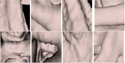
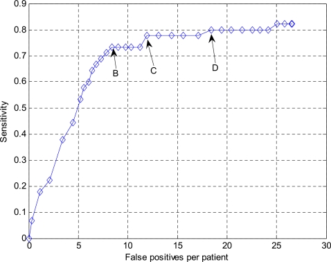
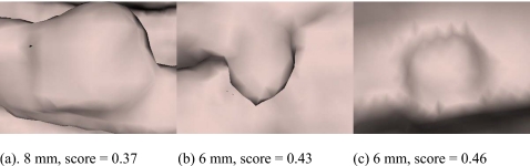
Computed tomographic colonography (CTC) computer aided detection (CAD) is a new method to detect colon polyps. Colonic polyps are abnormal growths that may become cancerous. Detection and removal of colonic polyps, particularly larger ones, has been shown to reduce the incidence of colorectal cancer. While high sensitivities and low false positive rates are consistently achieved for the detection of polyps sized 1 cm or larger, lower sensitivities and higher false positive rates occur when the goal of CAD is to identify "medium"-sized polyps, 6-9 mm in diameter. Such medium-sized polyps may be important for clinical patient management. We have developed a wavelet-based postprocessor to reduce false positives for this polyp size range. We applied the wavelet-based postprocessor to CTC CAD findings from 44 patients in whom 45 polyps with sizes of 6-9 mm were found at segmentally unblinded optical colonoscopy and visible on retrospective review of the CT colonography images. Prior to the application of the wavelet-based postprocessor, the CTC CAD system detected 33 of the polyps (sensitivity 73.33%) with 12.4 false positives per patient, a sensitivity comparable to that of expert radiologists. Fourfold cross validation with 5000 bootstraps showed that the wavelet-based postprocessor could reduce the false positives by 56.61% (p <0.001), to 5.38 per patient (95% confidence interval [4.41, 6.34]), without significant sensitivity degradation (32/45, 71.11%, 95% confidence interval [66.39%, 75.74%], p=0.1713). We conclude that this wavelet-based postprocessor can substantially reduce the false positive rate of our CTC CAD for this important polyp size range.
Figures


Similar articles
-
Computer-aided detection of colonic polyps at CT colonography using a Hessian matrix-based algorithm: preliminary study.AJR Am J Roentgenol. 2007 Jul;189(1):41-51. doi: 10.2214/AJR.07.2072. AJR Am J Roentgenol. 2007. PMID: 17579150
-
Mixture of expert 3D massive-training ANNs for reduction of multiple types of false positives in CAD for detection of polyps in CT colonography.Med Phys. 2008 Feb;35(2):694-703. doi: 10.1118/1.2829870. Med Phys. 2008. PMID: 18383691
-
Performance of a previously validated CT colonography computer-aided detection system in a new patient population.AJR Am J Roentgenol. 2008 Jul;191(1):168-74. doi: 10.2214/AJR.07.3354. AJR Am J Roentgenol. 2008. PMID: 18562741
-
CT colonography with computer-aided detection: recognizing the causes of false-positive reader results.Radiographics. 2014 Nov-Dec;34(7):1885-905. doi: 10.1148/rg.347130053. Radiographics. 2014. PMID: 25384290 Free PMC article. Review.
-
Computer-aided diagnosis for CT colonography.Semin Ultrasound CT MR. 2004 Oct;25(5):419-31. doi: 10.1053/j.sult.2004.07.002. Semin Ultrasound CT MR. 2004. PMID: 15559125 Review.
Cited by
-
Increasing computer-aided detection specificity by projection features for CT colonography.Med Phys. 2010 Apr;37(4):1468-81. doi: 10.1118/1.3302833. Med Phys. 2010. PMID: 20443468 Free PMC article.
-
EMPLOYING TOPOGRAPHICAL HEIGHT MAP IN COLONIC POLYP MEASUREMENT AND FALSE POSITIVE REDUCTION.Pattern Recognit. 2009;42(6):1029-1040. doi: 10.1016/j.patcog.2008.09.034. Pattern Recognit. 2009. PMID: 19578483 Free PMC article.
-
A review of computer-aided diagnosis in thoracic and colonic imaging.Quant Imaging Med Surg. 2012 Sep;2(3):163-76. doi: 10.3978/j.issn.2223-4292.2012.09.02. Quant Imaging Med Surg. 2012. PMID: 23256078 Free PMC article.
-
A CAD of fully automated colonic polyp detection for contrasted and non-contrasted CT scans.Int J Comput Assist Radiol Surg. 2017 Apr;12(4):627-644. doi: 10.1007/s11548-017-1521-9. Epub 2017 Jan 18. Int J Comput Assist Radiol Surg. 2017. PMID: 28101760
-
Optimizing computer-aided colonic polyp detection for CT colonography by evolving the Pareto fronta.Med Phys. 2009 Jan;36(1):201-12. doi: 10.1118/1.3040177. Med Phys. 2009. PMID: 19235388 Free PMC article.
References
Publication types
MeSH terms
Grants and funding
LinkOut - more resources
Full Text Sources
Other Literature Sources
Medical
Miscellaneous
