. 2009 Apr;126(4):514-22.
doi: 10.1111/j.1365-2567.2008.02913.x. Epub 2008 Sep 4.

Retinoic acid inhibits in vivo interleukin-2 gene expression and T-cell activation in mice

Affiliations

Retinoic acid inhibits in vivo interleukin-2 gene expression and T-cell activation in mice

Aase Ertesvag et al. Immunology. 2009 Apr.

Abstract

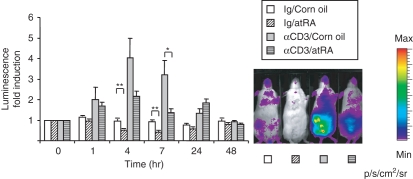
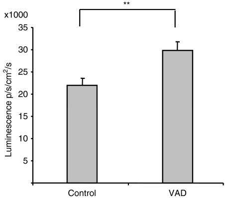
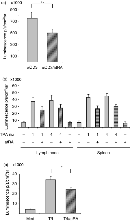
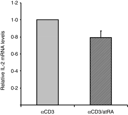
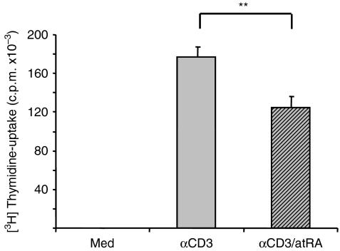
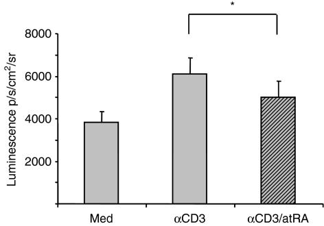
Interleukin-2 (IL-2) is an essential cytokine for T-lymphocyte homeostasis. We have previously reported that all-trans retinoic acid (atRA) enhances the secretion of IL-2 from human peripheral blood T cells in vitro, followed by increased proliferation and inhibition of spontaneous cell death. In this study we used a transgenic IL-2 gene luciferase reporter model to examine the effects of atRA in vivo. In contrast to the observations in human T cells, we found an overall reduction in luciferase-reported IL-2 gene expression in mice treated with atRA. Whole-body luminescence of anti-CD3-treated and non-treated mice was reduced in mice receiving atRA. Accordingly, after 7 hr, IL-2 gene expression was on average 55% lower in the atRA-treated mice compared with the control mice. Furthermore, mice fed a vitamin A-deficient diet had a significantly higher basal level of luciferase activity compared with control mice, demonstrating that vitamin A modulates IL-2 gene expression in vivo. Importantly, the atRA-mediated inhibition of IL-2 gene expression was accompanied by decreased DNA synthesis in murine T cells, suggesting a physiological relevance of the reduced IL-2 gene expression observed in transgenic reporter mice.


Figures

Figure 1
Figure 1
Down-regulation of interleukin-2 (IL-2) gene expression in whole mice following all-trans retinoic acid (atRA) administration. The IL-2 luciferase reporter mice were given atRA (50 mg/kg) or corn oil before injection of anti-CD3 antibody or control antibody (Ig) as indicated. Images were taken before atRA/anti-CD3 administration (0 hr) and then after 1 hr, 4 hr, 7 hr, 24 hr and 48 hr. (n= 5 at 24 hr and n= 3 at 48 hr, n= 10 at the other time-points). Average luciferase activity at each time-point is shown relative to luciferase activity at 0 hr ± SEM. *P< 0·05, **P< 0·01, independent samples t-test. One representative example of whole-body luminescence at 7 hr of mice treated as indicated is shown.
Figure 2
Figure 2
Vitamin A-deficient (VAD) mice have a higher basal activity of interleukin-2 (IL-2) gene expression. Twenty VAD mice and 20 age-matched controls were anaesthetized and injected with 200 μl luciferin in phosphate-buffered saline. After 7 min, luminescence of the whole mouse was examined. Average luminescence ± SEM is shown. **P< 0·01, n= 20, independent samples t-test.
Figure 3
Figure 3
All-trans retinoic acid (atRA) inhibits interleukin-2 (IL-2) gene activity in isolated cells. (a) T cells from spleen were cultures in triplicates with immobilized anti-CD3 (2 μg/ml) in the presence or absence of atRA (100 nm). (b) T cells from lymph nodes and spleen were cultured in triplicates with or without 12-O-tetradecanoylphorbol 13-acetate (TPA; 1 or 4 × 10⁻⁹m) in the presence or absence of atRA (100 nm). (c) T cells from spleen were cultured in triplicates with TPA (10−9 m) and ionomycin (0·5 μm) in the presence or absence of atRA (100 nm). (a–c) After 4 hr, luciferin was added, and luminescence was examined as described in the Materials and methods. Average luminescence ± SEM are shown of the median of triplicates from cells of nine mice (a and c) and four mice (b; **P< 0·01, paired samples t-test, *P< 0·05, Wilcoxon signed-rank test).
Figure 4
Figure 4
An antagonist of retinoic acid receptor (RAR) reverses the inhibiting effect of all-trans retinoic acid (atRA) on interleukin-2 (IL-2) expression. T cells from spleen were left untreated or stimulated with anti-CD3 (2 μg/ml) and anti-CD28 (1 μg/ml) in triplicates in the presence or absence of atRA (10 nm). Where indicated, cells were pretreated with Ro 41-5253 (500 nm). After 4 hr, luciferin was added, and the luminescence was examined. The average luminescence ± SEM of the median of triplicates from cells of six mice is shown, *P< 0·05, Wilcoxon signed-rank test.
Figure 5
Figure 5
All-trans retinoic acid (atRA) inhibits interleukin-2 (IL-2) messenger RNA (mRNA) in in vitro stimulated cells. T cells from spleen were stimulated with immobilized anti-CD3 (2 μg/ml) in the presence or absence of atRA (100 nm). After 4 hr cells were harvested and mRNA was isolated as described in the Materials and methods. Real time polymerase chain reaction was performed with primers to IL-2 and cyclophilin B was used as internal control. The average of relative IL-2 mRNA ± SEM is shown of cells of six mice.
Figure 6
Figure 6
All-trans retinoic acid (atRA) inhibits proliferation of isolated splenic T cells. T cells from spleen were left untreated or stimulated with immobilized anti-CD3 (2 μg/ml) in the presence or absence of atRA (100 nm) for 3 days. [3H]Thymidine was added for the last 24 hr, and DNA synthesis was determined as described in the Materials and methods. Bars represent average counts/min ± SEM of the median of triplicates of cells from eight mice. **P< 0·01, paired samples t-test.
Figure 7
Figure 7
All-trans retinoic acid (atRA) inhibits anti-CD3-stimulated nuclear factor-κB (NF-κB) activity in T cells. T cells were isolated from spleens of NF-κB-regulated luciferase reporter mice and cultured in triplicates alone or with immobilized anti-CD3 antibody (2 μg/ml) in the presence or absence of atRA (100 nm). After 4 hr, luminescence was recorded as described in the Materials and methods. The average luminescence of medians ± SEM (*P< 0·05, n= 5, Wilcoxon signed-rank test) is shown.

Similar articles

Cited by

References

    1. Gaffen SL, Liu KD. Overview of interleukin-2 function, production and clinical applications. Cytokine. 2004;28:109–23. - PubMed
    1. Rothenberg EV, Ward SB. A dynamic assembly of diverse transcription factors integrates activation and cell-type information for interleukin 2 gene regulation. Proc Natl Acad Sci USA. 1996;93:9358–65. - PMC - PubMed
    1. Zheng L, Fisher G, Miller RE, Peschon J, Lynch DH, Lenardo MJ. Induction of apoptosis in mature T cells by tumour necrosis factor. Nature. 1995;377:348–51. - PubMed
    1. Dhein J, Walczak H, Baumler C, Debatin KM, Krammer PH. Autocrine T-cell suicide mediated by APO-1/(Fas/CD95) Nature. 1995;373:438–41. - PubMed
    1. Hildeman DA, Zhu Y, Mitchell TC, Kappler J, Marrack P. Molecular mechanisms of activated T cell death in vivo. Curr Opin Immunol. 2002;14:354–9. - PubMed

Publication types

MeSH terms

LinkOut - more resources