Comparative Study
. 2008 Dec;7(6):836-49.
doi: 10.1111/j.1474-9726.2008.00430.x. Epub 2008 Sep 5.

Astrocytes from acyclic female rats exhibit lowered capacity for neuronal differentiation

Affiliations
Comparative Study

Astrocytes from acyclic female rats exhibit lowered capacity for neuronal differentiation

Danielle K Lewis et al. Aging Cell. 2008 Dec.

Abstract

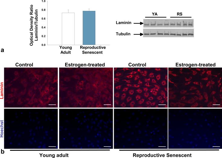
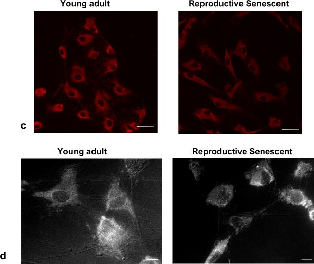
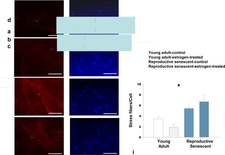
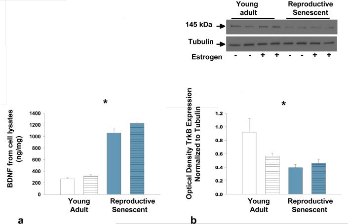
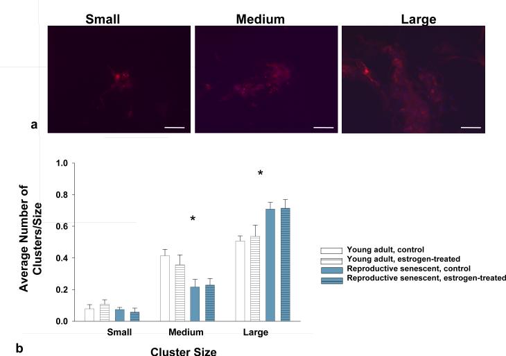
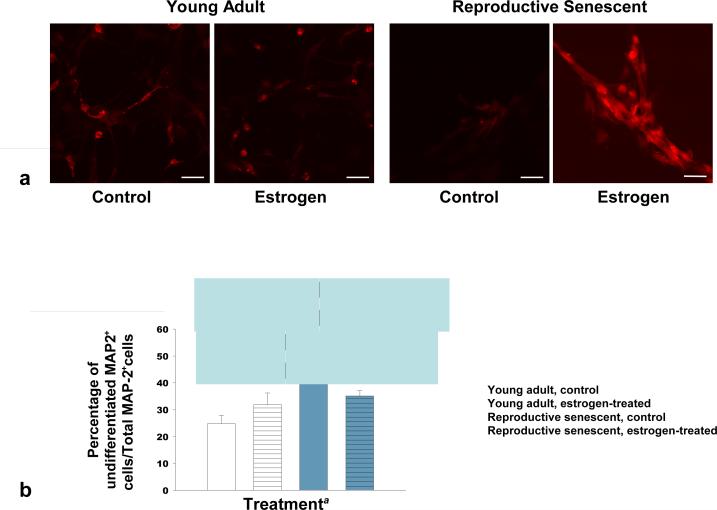
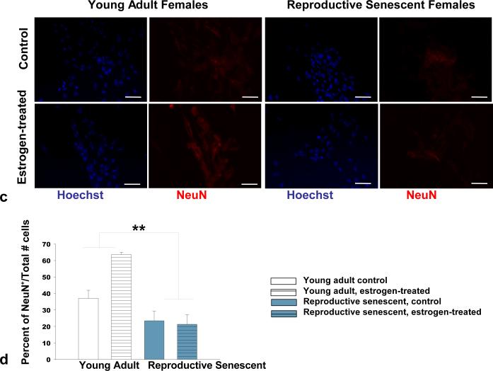
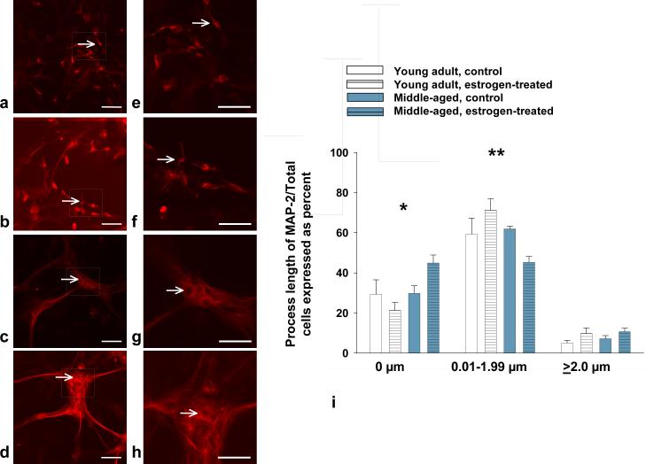
Astrocytes comprise a large proportion of the central nervous system support cells and play a critical role in neural injury and repair. The present study examined the impact of ovarian aging using an ex vivo model system, where astrocytes were derived from the olfactory bulb of young, reproductively competent females and reproductive senescent females. Cellular morphology and the spatial pattern of laminin deposition was altered in astrocyte cultures derived from reproductive senescent females. Young adult astrocytes had a flattened polygonal shape with actin bundles at the cell edges, while reproductive senescent astrocytes had a contractile appearance with thick stress fibers visible throughout the cell. Moreover, in reproductive senescent astrocytes, BDNF was elevated with a concomitant reduction in expression of the BDNF receptor, TrkB. To examine the ability of astrocytes derived from young adult and reproductive senescent females to promote neuronal differentiation, neural progenitor cells (NPCs) were co-cultured with astrocytes derived from these groups. At day 4 in vitro, MAP-2(+) NPCs were located in smaller clusters when co-cultured with young adult astrocytes and in large clusters when co-cultured with older astrocytes. At days 6 and 10, neuronal differentiation was significantly reduced in reproductive senescent astrocyte-NPC co-cultures, as determined by NeuN(+) cell numbers and MAP-2(+) process lengths. Furthermore, estrogen only enhanced neuronal differentiation in young adult-NPC co-cultures. The ovarian age-related astrocyte phenotype thus limits the ability of this cell to promote neuronal differentiation in NPC populations and suggests that the astrocyte-mediated microenvironment in older acyclic females is less conducive to repair following neurovascular injury.


Figures

FIG 1
FIG 1
Protein expression of laminin from young adult and reproductive senescent astrocytes. No differences in laminin protein expression were observed in young adult or reproductive senescent astrocytes by Western blot analysis (a). Laminin expression as examined by immunohistochemistry in young adult and reproductive senescent astrocytes displayed a different localization pattern (b,c). Astrocyte laminin expression (b, top panel) was analyzed in the presence or absence of estrogen (40 nM dose) and counter-stained with the nuclear dye Hoechst 33258 (b, bottom panel). A highly dispersed pattern of laminin deposition was seen in the young adult astrocyte cultures in contrast to the largely perinuclear concentration of laminin in reproductive senescent astrocyte cultures, both here and in 1c by confocal microscopy. Combined epifluorescence-phase contrast light microscopy (1d) indicates that laminin organization closely approximates the morphology of the cell. Bars represent mean ± SEM of a single representative experiment with n=4 for each treatment group. Bar: 50 μm.
FIG 1
FIG 1
Protein expression of laminin from young adult and reproductive senescent astrocytes. No differences in laminin protein expression were observed in young adult or reproductive senescent astrocytes by Western blot analysis (a). Laminin expression as examined by immunohistochemistry in young adult and reproductive senescent astrocytes displayed a different localization pattern (b,c). Astrocyte laminin expression (b, top panel) was analyzed in the presence or absence of estrogen (40 nM dose) and counter-stained with the nuclear dye Hoechst 33258 (b, bottom panel). A highly dispersed pattern of laminin deposition was seen in the young adult astrocyte cultures in contrast to the largely perinuclear concentration of laminin in reproductive senescent astrocyte cultures, both here and in 1c by confocal microscopy. Combined epifluorescence-phase contrast light microscopy (1d) indicates that laminin organization closely approximates the morphology of the cell. Bars represent mean ± SEM of a single representative experiment with n=4 for each treatment group. Bar: 50 μm.
FIG 2
FIG 2
F-actin stress fibers are significantly increased in reproductive senescent astrocytes as compared to young adult astrocytes. Stress fiber formation was analyzed in astrocytes cultured in the presence and absence of 40 nM estrogen by Alexa Fluor 594® phalloidin staining in young (a,b,i) and reproductive senescent astrocytes (c,d,i) and counter-stained with the nuclear dye Hoechst 33258 (e-h). Estrogen treatment attenuated stress fiber formation in young adults but not reproductive senescent astrocyte cultures. White arrow indicates presence of stress fibers. Bars represent mean ± SEM of a single representative experiment with n=4-5 for each treatment group. Interaction of age and hormone is statistically significant at p<0.05 and indicated by (*). Bar: 50 μm.
FIG 3
FIG 3
Protein expression of BDNF and TrkB in young adult and reproductive senescent astrocyte cell lysates. BDNF protein expression was determined by ELISA and TrkB expression was determined by immunoblotting. TrkB expression was normalized to α-tubulin. BDNF expression (a) increased with age while expression of TrkB (b) decreased with age in astrocyte cell lysates. Bars represent mean ± SEM of a single representative experiment with n=4-8 for each treatment group. Main effect of age was statistically significant at p<0.05 and is indicated by (*).
FIG 4
FIG 4
Neurosphere cluster size was used as a measure of putative neuronal maturity in NPCs co-cultured with young adult or reproductive senescent astrocytes (a). Small neurosphere clusters were defined as (a, far left) aggregates of NPCs consisting of 5-10 visible grouped nuclei, medium clusters (a, middle panel) as consisting of 11-20 visible grouped nuclei and large clusters (a, far right) as containing greater than 20 visible grouped nuclei. Cells were immunoreactive for MAP-2 (red, a) and counter-stained for the nuclear dye Hoechst 33258 (blue, a). The frequency of small cluster size did not differ between those cultured with young adult or reproductive senescent astrocytes (b). However, neurosphere aggregates co-cultured with young adult astrocytes had significantly more medium size clusters while reproductive senescent astrocyte co-cultures had predominantly large clusters (b). Bars represent mean ± SEM of a single representative experiment with n=5-6 for each treatment group. Statistical significance at p< 0.05 is indicated by (*). Bar: 50 μm.
FIG 5
FIG 5
Neuronal differentiation was significantly increased in co-cultures of NPCs with young adult astrocytes at day-6 as compared to co-cultures with reproductive senescent astrocytes (a-d). The percentage of non-process bearing MAP-2+ progenitor cells was increased in co-cultures with reproductive senescent astrocytes (a,b). Immunolabeling for the late stage neuronal marker, NeuN, was enhanced with estrogen only in young adult astrocyte:NPC co-cultures (c,d). Bars represent mean ± SEM of a single representative experiment with n=3-6 for each treatment group. Arrows indicate individual cell bodies. The main effect of age was statistically significant at p<0.05 and is indicated by (*). The interaction effect of age with estrogen exposure was statistically significant at p<0.05 and is indicated by (**). Bar: 50 μm.
FIG 5
FIG 5
Neuronal differentiation was significantly increased in co-cultures of NPCs with young adult astrocytes at day-6 as compared to co-cultures with reproductive senescent astrocytes (a-d). The percentage of non-process bearing MAP-2+ progenitor cells was increased in co-cultures with reproductive senescent astrocytes (a,b). Immunolabeling for the late stage neuronal marker, NeuN, was enhanced with estrogen only in young adult astrocyte:NPC co-cultures (c,d). Bars represent mean ± SEM of a single representative experiment with n=3-6 for each treatment group. Arrows indicate individual cell bodies. The main effect of age was statistically significant at p<0.05 and is indicated by (*). The interaction effect of age with estrogen exposure was statistically significant at p<0.05 and is indicated by (**). Bar: 50 μm.
FIG 6
FIG 6
Estrogen enhanced the length of MAP-2+ processes in NPCs co-cultured with young adult astrocytes (a,b,i) as compared to co-cultures with reproductive senescent astrocytes (c,d,i). Arrows indicate individual cell bodies. In the reproductive senescent astrocyte:NPC co-cultures, significantly more of non-process bearing cells were observed. Thus, the percentage of MAP-2+ cells with no visible processes was significantly increased in reproductive senescent astrocyte:NPC co-cultures and was exacerbated with estrogen treatment (i,*). Likewise, estrogen enhanced the percentage of MAP-2+ cells with processes between 0.01 and 1.99 μm in length in young adult astrocyte:NPC co-cultures as compared to co-cultures with reproductive senescent astrocytes (i, **). Boxed regions in image (a-d) are enlarged to show process detail for young adult (e,f) and reproductive senescent astrocytes (g,h). Bars represent mean ± SEM of a single representative experiment with n=4-6 for each treatment group. Interaction of age and hormone treatment was statistically significant at p<0.05 and is indicated by (*,**). Bar: 50 μm (a-d) or 20 μm (e-h).
FIG 7
FIG 7
DiI-Labeled PC12 cells co-cultured with young adult astrocytes for 6 days exhibited increased levels of TGFβ1 as compared to co-cultures with reproductive senescent astrocytes. NGF-induced neuronal differentiation of PC12 cells significantly increased the protein expression of TGFβ1 (a). An increase in TGFβ1 protein expression was also observed when PC12 cells were co-cultured with young adult astrocytes but not when PC12 co-cultured with reproductive senescent astrocytes or in astrocyte only cultures (b). 0 indicates that no detectable level of TGFβ1 was observed. Bars represent mean ± SEM of a single representative experiment with n=3-5 for each treatment group. Statistical significance at p< 0.05 is indicated by (*, **). Bar: 50 μm.

Similar articles

Cited by

References

    1. Amateau SK, McCarthy MM. Sexual differentiation of astrocyte morphology in the developing rat preoptic area. J. Neuroendocrinol. 2002;14:904–910. - PubMed
    1. Bailly K, Ridley AJ, Hall SM, Haworth SG. RhoA activation by hypoxia in pulmonary arterial smooth muscle cells is age and site specific. Circ. Res. 2004;94:1383–1391. - PubMed
    1. Bake S, Sohrabji F. 17β-Estradiol differentially regulate blood-brain barrier permeability in young and aging female rats. Endocrinol. 2004;145:5471–5475. - PubMed
    1. Banner LR, Moayeri NN, Patterson PH. Leukemia inhibitory factor is expressed in astrocytes following cortical brain injury. Exp. Neurol. 1997;147:1–9. - PubMed
    1. Camarillo C, Kumar LS, Bake S, Sohrabji F, Miranda RC. Ethanol regulates angiogenic cytokines during neural development: evidence from an in vitro model of mitogenwithdrawal-induced cerebral cortical neuroepithelial differentiation. Alcohol Clin. Exp. Res. 2007;31:324–335. - PMC - PubMed

Publication types